Back


Poster Session D - Tuesday Morning
Category: General Endoscopy
D0293 - Outside In: Endometriosis of the Appendix, Cecum, and Ileum Masquerading as Suspected Crohn’s Disease
Tuesday, October 25, 2022
10:00 AM – 12:00 PM ET
Location: Crown Ballroom

Has Audio

Jonathan J. Cho, MD, PhD
Naval Medical Center San Diego
San Diego, CA
Presenting Author(s)
Award: Presidential Poster Award
Jonathan J. Cho, MD, PhD1, Kevin Pak, MD1, Ryan P. Austin, MD2, Leybelis Padilla, MD1, Rashad C. Wilkerson, DO1, Theodore D. Edson, MD2
1Naval Medical Center San Diego, San Diego, CA; 2Naval Hospital Camp Pendleton, Oceanside, CA
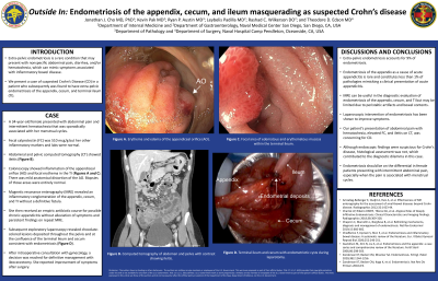
Introduction: Extra-pelvic endometriosis is a rare condition that may present with nonspecific abdominal pain, diarrhea, and/or hematochezia, which can mimic symptoms associated with inflammatory bowel disease. We present a case of suspected Crohn’s Disease (CD) in a patient who subsequently was found to have extra-pelvic endometriosis of the appendix, cecum, and terminal ileum (TI).
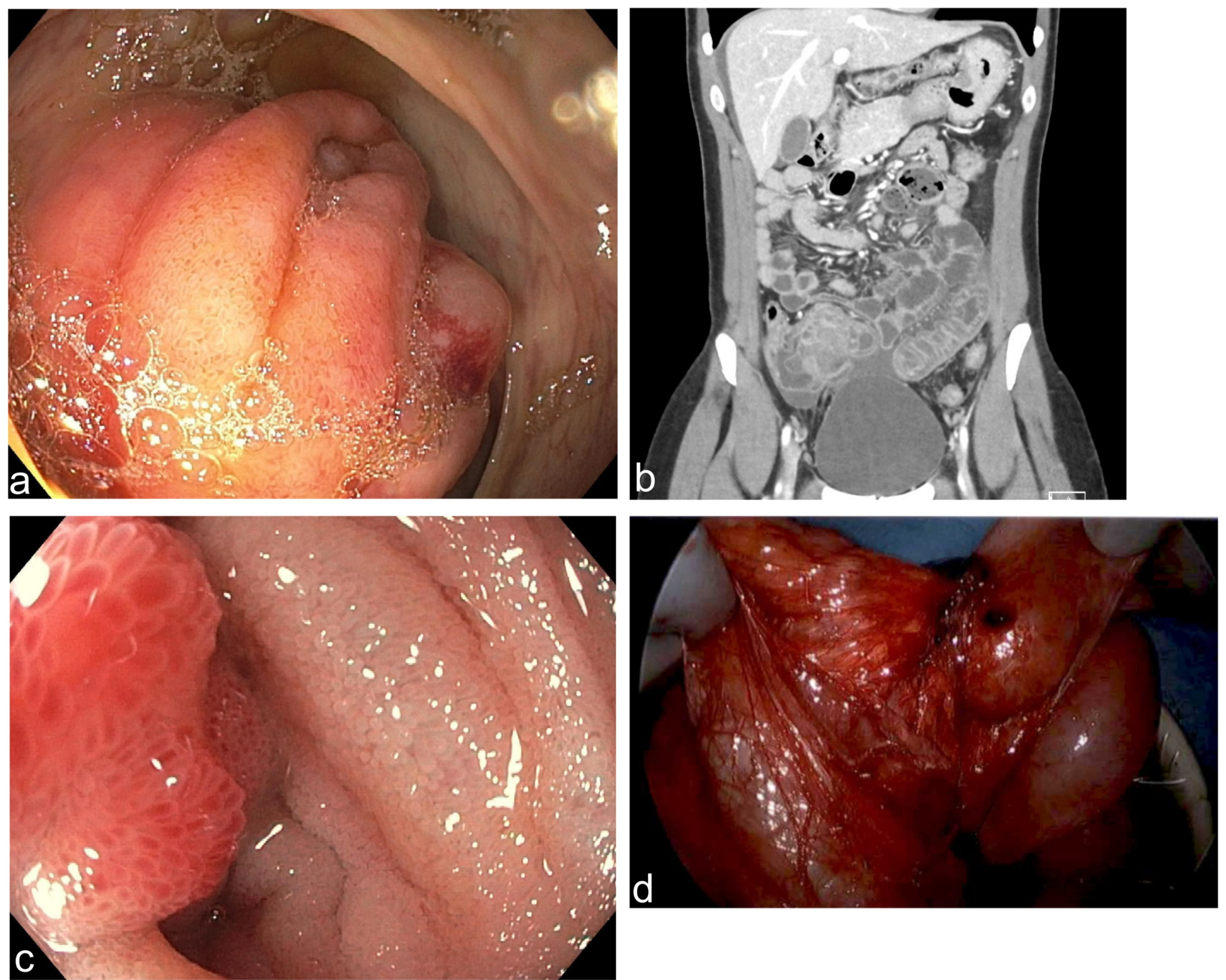
Case Description/Methods: A 34-year-old female presented with abdominal pain and intermittent hematochezia that was sporadically associated with her menstrual cycles. Her fecal calprotectin (FC) was 510 mcg/g but her other inflammatory markers and labs were normal. Abdominal and pelvic computed tomography (CT) showed ileitis. Index colonoscopy showed inflammation of the appendiceal orifice and focal erythema in the TI. Biopsies of those areas were entirely normal. Magnetic resonance enterography (MRE) revealed an inflammatory conglomeration of the appendix, cecum, and TI without a definitive fistula. Repeat colonoscopy showed similar findings with mild architectural distortion of the appendiceal orifice. She then received an empiric antibiotic course for possible chronic appendicitis without alleviation of symptoms and persistent findings on repeat MRE. Subsequent exploratory laparoscopy revealed chocolate-colored lesions deposited throughout the pelvis and at the confluence of the terminal ileum and cecum consistent with endometriosis. After intraoperative consultation with gynecology, a decision was reached for definitive management with ileocecectomy. She reported improvement of symptoms after surgery.
Discussion: Extra-pelvic endometriosis accounts for 9% of endometriosis. Endometriosis of the appendix as a cause of acute appendicitis is rare and constitutes less than 1% of pathologies mimicking a clinical presentation of acute appendicitis. MRE can be useful in the diagnostic evaluation of endometriosis of the appendix, cecum, and TI but may be limited due to peristaltic artifacts and bowel contents. Laparoscopic intervention of endometriosis has been shown to improve symptoms. Our patient’s presentation of abdominal pain with hematochezia, elevated FC, and ileitis on CT, was concerning for CD. Although endoscopic findings were suspicious for Crohn’s disease, histological assessment was not, which contributed to the diagnostic dilemma in this case. Endometriosis should be on the differential in female patients presenting with intermittent abdominal pain, especially when the pain is associated with menstrual cycles.

Disclosures:
Jonathan J. Cho, MD, PhD1, Kevin Pak, MD1, Ryan P. Austin, MD2, Leybelis Padilla, MD1, Rashad C. Wilkerson, DO1, Theodore D. Edson, MD2. D0293 - Outside In: Endometriosis of the Appendix, Cecum, and Ileum Masquerading as Suspected Crohn’s Disease, ACG 2022 Annual Scientific Meeting Abstracts. Charlotte, NC: American College of Gastroenterology.
Jonathan J. Cho, MD, PhD1, Kevin Pak, MD1, Ryan P. Austin, MD2, Leybelis Padilla, MD1, Rashad C. Wilkerson, DO1, Theodore D. Edson, MD2
1Naval Medical Center San Diego, San Diego, CA; 2Naval Hospital Camp Pendleton, Oceanside, CA
Introduction: Extra-pelvic endometriosis is a rare condition that may present with nonspecific abdominal pain, diarrhea, and/or hematochezia, which can mimic symptoms associated with inflammatory bowel disease. We present a case of suspected Crohn’s Disease (CD) in a patient who subsequently was found to have extra-pelvic endometriosis of the appendix, cecum, and terminal ileum (TI).
Case Description/Methods: A 34-year-old female presented with abdominal pain and intermittent hematochezia that was sporadically associated with her menstrual cycles. Her fecal calprotectin (FC) was 510 mcg/g but her other inflammatory markers and labs were normal. Abdominal and pelvic computed tomography (CT) showed ileitis. Index colonoscopy showed inflammation of the appendiceal orifice and focal erythema in the TI. Biopsies of those areas were entirely normal. Magnetic resonance enterography (MRE) revealed an inflammatory conglomeration of the appendix, cecum, and TI without a definitive fistula. Repeat colonoscopy showed similar findings with mild architectural distortion of the appendiceal orifice. She then received an empiric antibiotic course for possible chronic appendicitis without alleviation of symptoms and persistent findings on repeat MRE. Subsequent exploratory laparoscopy revealed chocolate-colored lesions deposited throughout the pelvis and at the confluence of the terminal ileum and cecum consistent with endometriosis. After intraoperative consultation with gynecology, a decision was reached for definitive management with ileocecectomy. She reported improvement of symptoms after surgery.
Discussion: Extra-pelvic endometriosis accounts for 9% of endometriosis. Endometriosis of the appendix as a cause of acute appendicitis is rare and constitutes less than 1% of pathologies mimicking a clinical presentation of acute appendicitis. MRE can be useful in the diagnostic evaluation of endometriosis of the appendix, cecum, and TI but may be limited due to peristaltic artifacts and bowel contents. Laparoscopic intervention of endometriosis has been shown to improve symptoms. Our patient’s presentation of abdominal pain with hematochezia, elevated FC, and ileitis on CT, was concerning for CD. Although endoscopic findings were suspicious for Crohn’s disease, histological assessment was not, which contributed to the diagnostic dilemma in this case. Endometriosis should be on the differential in female patients presenting with intermittent abdominal pain, especially when the pain is associated with menstrual cycles.

Figure: Figure 1. Endometriosis of the appendix, cecum, and terminal ileum. a, Erythema and edema of the appendiceal orifice on first colonoscopy. b, Computed tomography of abdomen and pelvis with contrast showing ileitis. c, Focal area of edematous and erythematous mucosa within the terminal ileum on second colonoscopy. d, Terminal ileum and cecum with endometriotic cysts during laparotomy.
Disclosures:
Jonathan Cho indicated no relevant financial relationships.
Kevin Pak indicated no relevant financial relationships.
Ryan Austin indicated no relevant financial relationships.
Leybelis Padilla indicated no relevant financial relationships.
Rashad Wilkerson indicated no relevant financial relationships.
Theodore Edson indicated no relevant financial relationships.
Jonathan J. Cho, MD, PhD1, Kevin Pak, MD1, Ryan P. Austin, MD2, Leybelis Padilla, MD1, Rashad C. Wilkerson, DO1, Theodore D. Edson, MD2. D0293 - Outside In: Endometriosis of the Appendix, Cecum, and Ileum Masquerading as Suspected Crohn’s Disease, ACG 2022 Annual Scientific Meeting Abstracts. Charlotte, NC: American College of Gastroenterology.

