Back


Poster Session D - Tuesday Morning
Category: GI Bleeding
D0321 - Hemorrhagic Shock as a Complication of Meckel's Diverticular Bleed
Tuesday, October 25, 2022
10:00 AM – 12:00 PM ET
Location: Crown Ballroom

Has Audio

Maaz Arif, MD
Wright State University
Granger, IN
Presenting Author(s)
Maaz Arif, MD1, We'am Hussain, MD2, Drew Triplett, DO1
1Wright State University, Dayton, OH; 2Wright State University Boonshoft School of Medicine, Dayton, OH
Introduction: Meckel’s diverticulum (MD) is the most common congenital abnormality of the gastrointestinal tract resulting from a failure of vitelline duct obliteration. While often presenting with painless hematochezia in children, MD can less frequently occur in adults but with severe and rare complications, including obstruction, perforation, or hemorrhage. We present a case of an 18-year-old male who presents with hemorrhagic shock from a MD bleed.
Case Description/Methods: A previously healthy 18-year-old male initially presented to an outside children’s hospital with abdominal pain and large volume hematochezia. Physical evaluation revealed orthostatic instability, tachycardia, hypotension, with a documented 900mL blood loss from the rectum. Hemoglobin level reached as low as 7.1 g/dL prior to receiving 6 units of packed red blood cells, fluids, and initiation of vasopressor support. After medical stabilization, the patient was transferred to our adult facility for higher acuity management. It is noteworthy that the patient presented to the same children’s hospital with similar, albeit milder, symptomatology five days prior and colonoscopy and esophagogastroduodenoscopy at the time were nonrevealing.
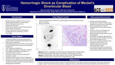
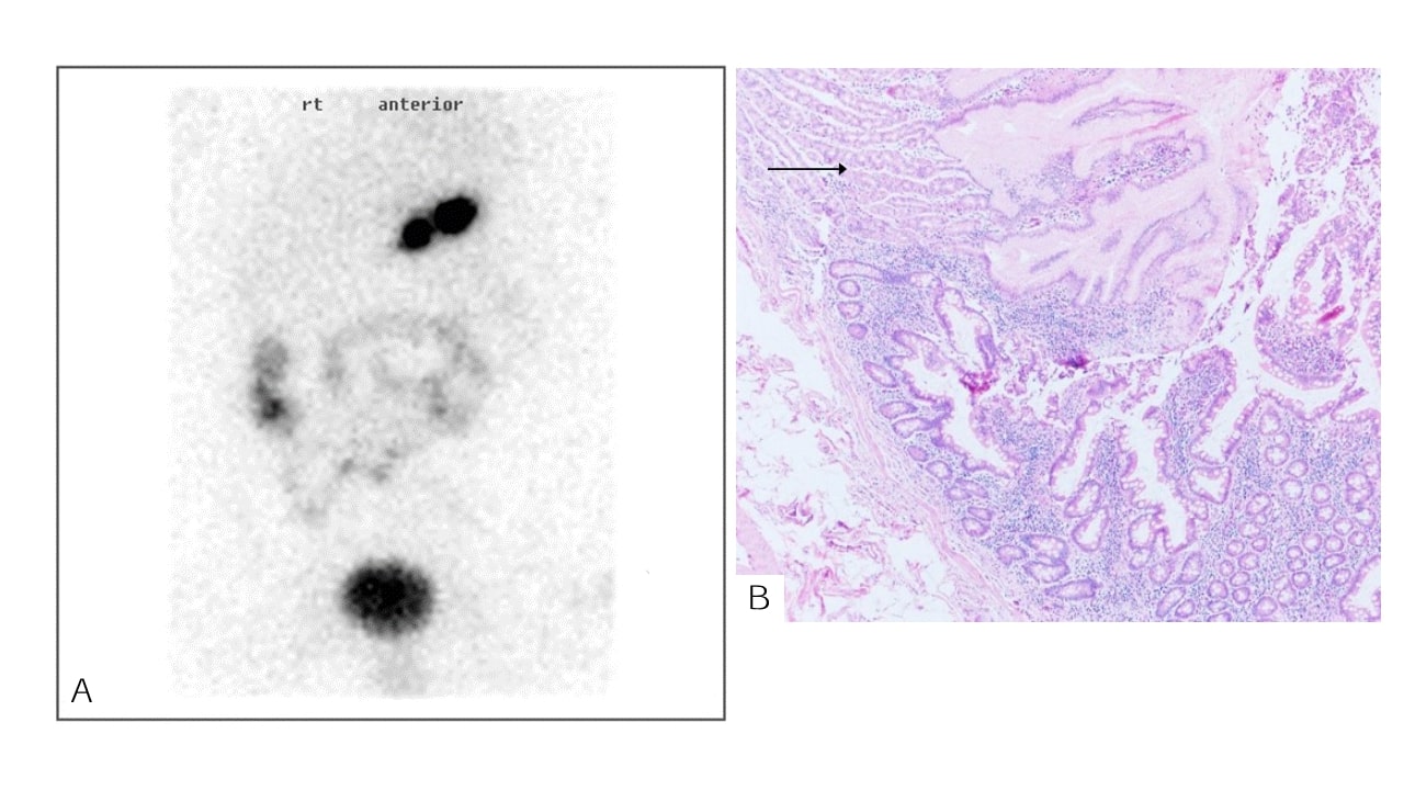
Our CT angiogram did not identify any foci of bleeding. A Meckel’s scan (Figure 1A) was obtained to further elucidate etiology, which was positive and revealed a solitary focus of increased uptake located within the right lower abdomen. The patient was transferred back to the children’s hospital for laparoscopic diverticulectomy. The surgically resected specimen revealed focal ulceration and pathology confirming gastric heterotropia (Figure 1B). The patient’s clinical recovery was subsequently uneventful.
Discussion: Meckel’s diverticulum typically presents with painless hematochezia in children, but its presentation can be more severe in the adult population. In fact, complications include obstruction (36.5%), intussusception (13.7%), and hemorrhage (11.8%). As in our case, we suspected the diagnosis given hemorrhage in a healthy young male preceded by an otherwise negative workup as neither upper gastrointestinal endoscopy, colonoscopy, or CT angiography were able to localize the source of bleeding. The diverticulum causes acidic secretions from embedded gastric tissue resulting in ulceration and hemorrhage of adjacent small bowel. Our case was diagnosed radiologically by the 99-mTc-pertechnetate Meckel’s scan because of the tracer’s affinity to the gastric mucosa, and has a sensitivity of 60% in adults.

Disclosures:
Maaz Arif, MD1, We'am Hussain, MD2, Drew Triplett, DO1. D0321 - Hemorrhagic Shock as a Complication of Meckel's Diverticular Bleed, ACG 2022 Annual Scientific Meeting Abstracts. Charlotte, NC: American College of Gastroenterology.
1Wright State University, Dayton, OH; 2Wright State University Boonshoft School of Medicine, Dayton, OH
Introduction: Meckel’s diverticulum (MD) is the most common congenital abnormality of the gastrointestinal tract resulting from a failure of vitelline duct obliteration. While often presenting with painless hematochezia in children, MD can less frequently occur in adults but with severe and rare complications, including obstruction, perforation, or hemorrhage. We present a case of an 18-year-old male who presents with hemorrhagic shock from a MD bleed.
Case Description/Methods: A previously healthy 18-year-old male initially presented to an outside children’s hospital with abdominal pain and large volume hematochezia. Physical evaluation revealed orthostatic instability, tachycardia, hypotension, with a documented 900mL blood loss from the rectum. Hemoglobin level reached as low as 7.1 g/dL prior to receiving 6 units of packed red blood cells, fluids, and initiation of vasopressor support. After medical stabilization, the patient was transferred to our adult facility for higher acuity management. It is noteworthy that the patient presented to the same children’s hospital with similar, albeit milder, symptomatology five days prior and colonoscopy and esophagogastroduodenoscopy at the time were nonrevealing.
Our CT angiogram did not identify any foci of bleeding. A Meckel’s scan (Figure 1A) was obtained to further elucidate etiology, which was positive and revealed a solitary focus of increased uptake located within the right lower abdomen. The patient was transferred back to the children’s hospital for laparoscopic diverticulectomy. The surgically resected specimen revealed focal ulceration and pathology confirming gastric heterotropia (Figure 1B). The patient’s clinical recovery was subsequently uneventful.
Discussion: Meckel’s diverticulum typically presents with painless hematochezia in children, but its presentation can be more severe in the adult population. In fact, complications include obstruction (36.5%), intussusception (13.7%), and hemorrhage (11.8%). As in our case, we suspected the diagnosis given hemorrhage in a healthy young male preceded by an otherwise negative workup as neither upper gastrointestinal endoscopy, colonoscopy, or CT angiography were able to localize the source of bleeding. The diverticulum causes acidic secretions from embedded gastric tissue resulting in ulceration and hemorrhage of adjacent small bowel. Our case was diagnosed radiologically by the 99-mTc-pertechnetate Meckel’s scan because of the tracer’s affinity to the gastric mucosa, and has a sensitivity of 60% in adults.

Figure: A. 99-mTc-pertechnetate scintigraphy image revealing a focal "hot spot" of increased uptake in the patient's right lower abdomen
B. Histopathology shows gastric tissue (arrow) embedded within adjacent normal small bowel tissue in the bottom half of the image (H&E stain; ×40)
B. Histopathology shows gastric tissue (arrow) embedded within adjacent normal small bowel tissue in the bottom half of the image (H&E stain; ×40)
Disclosures:
Maaz Arif indicated no relevant financial relationships.
We'am Hussain indicated no relevant financial relationships.
Drew Triplett indicated no relevant financial relationships.
Maaz Arif, MD1, We'am Hussain, MD2, Drew Triplett, DO1. D0321 - Hemorrhagic Shock as a Complication of Meckel's Diverticular Bleed, ACG 2022 Annual Scientific Meeting Abstracts. Charlotte, NC: American College of Gastroenterology.
