Back


Poster Session B - Monday Morning
Category: GI Bleeding
B0337 - Duodenal Hemorrhage From Eroding Pancreatic Metastases of Renal Cell Carcinoma
Monday, October 24, 2022
10:00 AM – 12:00 PM ET
Location: Crown Ballroom

Has Audio

Ameya Deshmukh, DO
Saint Louis University School of Medicine
St. Louis, MO
Presenting Author(s)
Ameya Deshmukh, DO1, Zarir Ahmed, DO2, Michelle Baliss, DO2, Jason Taylor, MD2, Antonio Cheesman, MD2
1Saint Louis University School of Medicine, St. Louis, MO; 2Saint Louis University, St. Louis, MO
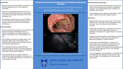
Introduction: Clear cell renal cell carcinoma (RCC) comprises 3% of all adult malignancies and is often diagnosed incidentally. Metachronous metastatic disease can occur even several years after nephrectomy with curative intent. Pancreatic and duodenal metastases from RCC are exceedingly rare. We describe an unusual case of upper GI bleeding from pancreatic RCC metastases eroding into the duodenum, diagnosed over 2 decades following nephrectomy.
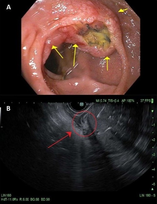
Case Description/Methods: A 62-year-old male with a history of clear cell RCC and distant nephrectomy (1999) believed to be in remission, presented to an outside hospital with melena, hemorrhagic shock, and hemoglobin of 5.9 g/dL. EGD revealed a protuberant ulcerated lesion in the post-bulbar region of the second portion of the duodenum with stigmata of recent bleeding, treated with hemostatic clip placement. Due to bleeding recurrence, repeat EGD with epinephrine injection and APC was done but failed to achieve hemostasis, as did subsequent embolization of the GDA by interventional radiology. He was transferred to our institution where he underwent successful embolization of the celiac and superior mesenteric artery branches. CT showed a 4.8 cm pancreatic head mass. Follow-up EGD with EUS demonstrated a hypervascular mass in the pancreatic head eroding into the adjacent duodenum. Fine needle biopsies were consistent with metastatic clear cell RCC. Further staging work-up showed no other lesions, and the patient underwent successful Whipple resection.
Discussion: Metastatic RCC outcomes are poor with 1-year survival less than 50%. Usual sites of RCC metastasis are lung, soft tissue, bone, and rarely, the small intestine or pancreas. Interestingly, pancreatic RCC metastases are frequently found as the only site of metastasis and thus can potentially be managed with surgical resection to improve prognosis. Therefore, prompt recognition and identification of disease extent with EUS and cross-sectional imaging aids in determining feasibility of surgical resection. Unfortunately, early identification is hindered by the asymptomatic nature of pancreatic metastases. In this case, erosion of the pancreatic metastases into the duodenum resulted in life-threatening hemorrhage that led to the appropriate diagnostic work-up, and ultimately, surgical management. This case highlights a rare presentation of metastatic RCC and emphasizes the importance of maintaining a high index of suspicion for metastatic RCC even years after nephrectomy.

Disclosures:
Ameya Deshmukh, DO1, Zarir Ahmed, DO2, Michelle Baliss, DO2, Jason Taylor, MD2, Antonio Cheesman, MD2. B0337 - Duodenal Hemorrhage From Eroding Pancreatic Metastases of Renal Cell Carcinoma, ACG 2022 Annual Scientific Meeting Abstracts. Charlotte, NC: American College of Gastroenterology.
1Saint Louis University School of Medicine, St. Louis, MO; 2Saint Louis University, St. Louis, MO
Introduction: Clear cell renal cell carcinoma (RCC) comprises 3% of all adult malignancies and is often diagnosed incidentally. Metachronous metastatic disease can occur even several years after nephrectomy with curative intent. Pancreatic and duodenal metastases from RCC are exceedingly rare. We describe an unusual case of upper GI bleeding from pancreatic RCC metastases eroding into the duodenum, diagnosed over 2 decades following nephrectomy.
Case Description/Methods: A 62-year-old male with a history of clear cell RCC and distant nephrectomy (1999) believed to be in remission, presented to an outside hospital with melena, hemorrhagic shock, and hemoglobin of 5.9 g/dL. EGD revealed a protuberant ulcerated lesion in the post-bulbar region of the second portion of the duodenum with stigmata of recent bleeding, treated with hemostatic clip placement. Due to bleeding recurrence, repeat EGD with epinephrine injection and APC was done but failed to achieve hemostasis, as did subsequent embolization of the GDA by interventional radiology. He was transferred to our institution where he underwent successful embolization of the celiac and superior mesenteric artery branches. CT showed a 4.8 cm pancreatic head mass. Follow-up EGD with EUS demonstrated a hypervascular mass in the pancreatic head eroding into the adjacent duodenum. Fine needle biopsies were consistent with metastatic clear cell RCC. Further staging work-up showed no other lesions, and the patient underwent successful Whipple resection.
Discussion: Metastatic RCC outcomes are poor with 1-year survival less than 50%. Usual sites of RCC metastasis are lung, soft tissue, bone, and rarely, the small intestine or pancreas. Interestingly, pancreatic RCC metastases are frequently found as the only site of metastasis and thus can potentially be managed with surgical resection to improve prognosis. Therefore, prompt recognition and identification of disease extent with EUS and cross-sectional imaging aids in determining feasibility of surgical resection. Unfortunately, early identification is hindered by the asymptomatic nature of pancreatic metastases. In this case, erosion of the pancreatic metastases into the duodenum resulted in life-threatening hemorrhage that led to the appropriate diagnostic work-up, and ultimately, surgical management. This case highlights a rare presentation of metastatic RCC and emphasizes the importance of maintaining a high index of suspicion for metastatic RCC even years after nephrectomy.

Figure: Figure 1: Eroding ulcerated mass in the duodenum seen on EGD (A). Pancreatic mass seen on EUS (B).
Disclosures:
Ameya Deshmukh indicated no relevant financial relationships.
Zarir Ahmed indicated no relevant financial relationships.
Michelle Baliss indicated no relevant financial relationships.
Jason Taylor indicated no relevant financial relationships.
Antonio Cheesman indicated no relevant financial relationships.
Ameya Deshmukh, DO1, Zarir Ahmed, DO2, Michelle Baliss, DO2, Jason Taylor, MD2, Antonio Cheesman, MD2. B0337 - Duodenal Hemorrhage From Eroding Pancreatic Metastases of Renal Cell Carcinoma, ACG 2022 Annual Scientific Meeting Abstracts. Charlotte, NC: American College of Gastroenterology.
