Back


Poster Session C - Monday Afternoon
Category: Biliary/Pancreas
C0081 - EBV-Positive Fibroblastic Reticular Cell Tumor of the Spleen With Pancreatic Invasion
Monday, October 24, 2022
3:00 PM – 5:00 PM ET
Location: Crown Ballroom

Has Audio

Syed Hamaad Rahman, DO
Methodist Dallas Medical Center
Dallas, TX
Presenting Author(s)
Syed Hamaad Rahman, DO1, Ali Waqar Chaudhry, MD2, Sadaf Raoof, MD3, Baha Aldeen Bani Fawwaz, MBBS3, Aimen Farooq, MD3, Abu Hurairah, MD3, Raphael Itzkowitz, DO4, Jie Ouyang, MD3
1Methodist Dallas Medical Center, Dallas, TX; 2FMH College of Medicine & Dentistry, Lahore, Punjab, Pakistan; 3AdventHealth Orlando, Orlando, FL; 4AdventHealth, Oviedo, FL
Introduction: Fibroblastic reticulum cells (FBRCs) are a subtype of dendritic cells, which are stromal cells found in structures such as the spleen and tonsils. They are accessory cells of the immune system with structural and functional roles. Other subtypes of dendritic cells include follicular dendritic cells (FDC) and interdigitating dendritic cells (IDC). Tumors arising from FDCs and IDCs are common, whereas tumors arising from FBRCs are extremely rare. To our knowledge, this is the only documented case of Epstein-Barr virus (EBV) positive FBRC tumor of splenic origin.
Case Description/Methods: A 37-year-old male presented with a 1-week history of worsening left upper quadrant abdominal pain. The pain was associated with night sweats and was worse with inspiration and palpation. He also reported increased weakness, fatigue, and loss of appetite.
A contrast CT of the abdomen and pelvis revealed a large, calcified heterogeneous splenic mass with lobulated contours and ill-defined margins inferiorly, concerning for neoplastic process with metastases. PET scan demonstrated two additional splenic lesions, one at the apical posterior aspect and another at the inferior margin.
The patient underwent splenectomy and open distal pancreatectomy as the splenic mass was found to be invading the distal pancreas. Histopathological analysis demonstrated a well circumscribed tumor within splenic parenchyma composed of spindle cells with atypical, vesicular ovoid nuclei with pale eosinophilic cytoplasm arranged in a fascicular fashion with a background of chronic inflammatory infiltrate. Immunohistochemistry analysis showed these cells were positive for SMA and negative for pan-keratin, CAM5.2, desmin, S100 protein, CD21, CD34, and CD35. The spindle cells displayed nuclear positivity for EBV-encoded RNA by in-situ hybridization. A diagnosis of EBV positive FBRC tumor was established.
Discussion: According to our review of the literature, there are only two documented cases of FBRC tumors arising from the spleen, neither of which was EBV positive. Therefore, our case is the first documented case of EBV positive FBRC of splenic origin. They are often mistaken as metastatic lesions and are grossly indistinguishable from other splenic malignancies. Microscopic analysis is critical for accurately diagnosing this entity. It is thought to be an indolent and benign lesion, but the malignant potential is not clear given lack of literature. Further research is needed to investigate the role of EBV in development of this disease.

Disclosures:
Syed Hamaad Rahman, DO1, Ali Waqar Chaudhry, MD2, Sadaf Raoof, MD3, Baha Aldeen Bani Fawwaz, MBBS3, Aimen Farooq, MD3, Abu Hurairah, MD3, Raphael Itzkowitz, DO4, Jie Ouyang, MD3. C0081 - EBV-Positive Fibroblastic Reticular Cell Tumor of the Spleen With Pancreatic Invasion, ACG 2022 Annual Scientific Meeting Abstracts. Charlotte, NC: American College of Gastroenterology.
1Methodist Dallas Medical Center, Dallas, TX; 2FMH College of Medicine & Dentistry, Lahore, Punjab, Pakistan; 3AdventHealth Orlando, Orlando, FL; 4AdventHealth, Oviedo, FL
Introduction: Fibroblastic reticulum cells (FBRCs) are a subtype of dendritic cells, which are stromal cells found in structures such as the spleen and tonsils. They are accessory cells of the immune system with structural and functional roles. Other subtypes of dendritic cells include follicular dendritic cells (FDC) and interdigitating dendritic cells (IDC). Tumors arising from FDCs and IDCs are common, whereas tumors arising from FBRCs are extremely rare. To our knowledge, this is the only documented case of Epstein-Barr virus (EBV) positive FBRC tumor of splenic origin.
Case Description/Methods: A 37-year-old male presented with a 1-week history of worsening left upper quadrant abdominal pain. The pain was associated with night sweats and was worse with inspiration and palpation. He also reported increased weakness, fatigue, and loss of appetite.
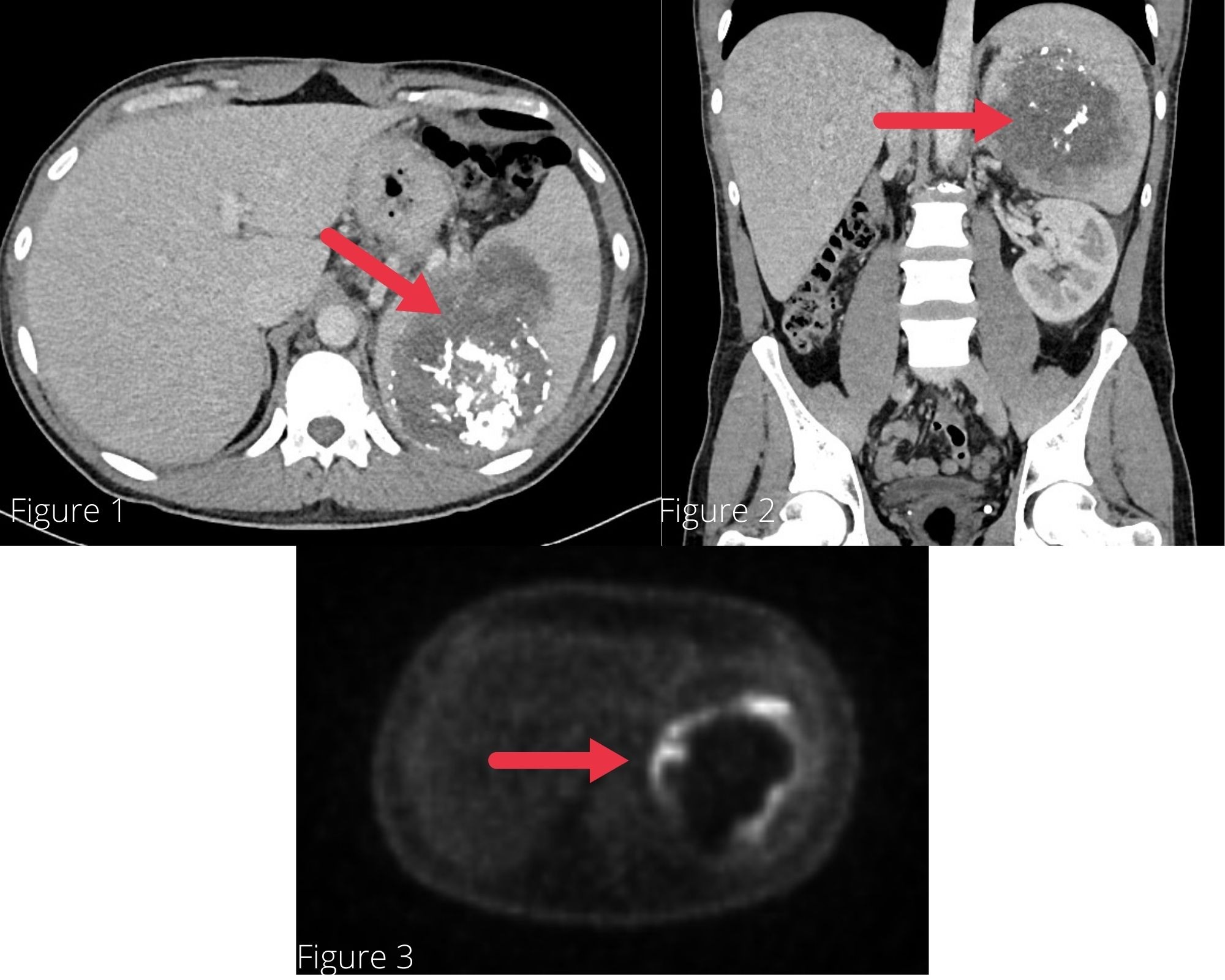
A contrast CT of the abdomen and pelvis revealed a large, calcified heterogeneous splenic mass with lobulated contours and ill-defined margins inferiorly, concerning for neoplastic process with metastases. PET scan demonstrated two additional splenic lesions, one at the apical posterior aspect and another at the inferior margin.
The patient underwent splenectomy and open distal pancreatectomy as the splenic mass was found to be invading the distal pancreas. Histopathological analysis demonstrated a well circumscribed tumor within splenic parenchyma composed of spindle cells with atypical, vesicular ovoid nuclei with pale eosinophilic cytoplasm arranged in a fascicular fashion with a background of chronic inflammatory infiltrate. Immunohistochemistry analysis showed these cells were positive for SMA and negative for pan-keratin, CAM5.2, desmin, S100 protein, CD21, CD34, and CD35. The spindle cells displayed nuclear positivity for EBV-encoded RNA by in-situ hybridization. A diagnosis of EBV positive FBRC tumor was established.
Discussion: According to our review of the literature, there are only two documented cases of FBRC tumors arising from the spleen, neither of which was EBV positive. Therefore, our case is the first documented case of EBV positive FBRC of splenic origin. They are often mistaken as metastatic lesions and are grossly indistinguishable from other splenic malignancies. Microscopic analysis is critical for accurately diagnosing this entity. It is thought to be an indolent and benign lesion, but the malignant potential is not clear given lack of literature. Further research is needed to investigate the role of EBV in development of this disease.

Figure: Figure 1 and 2- CT abdomen showing a large heterogenous splenic mass with lobulated contours and irregular calcifications concerning for neoplastic process. Figure 3- PET scan showing a large heterogeneous splenic mass with rim of severe hypermetabolism consistent with a malignancy.
Disclosures:
Syed Hamaad Rahman indicated no relevant financial relationships.
Ali Waqar Chaudhry indicated no relevant financial relationships.
Sadaf Raoof indicated no relevant financial relationships.
Baha Aldeen Bani Fawwaz indicated no relevant financial relationships.
Aimen Farooq indicated no relevant financial relationships.
Abu Hurairah indicated no relevant financial relationships.
Raphael Itzkowitz indicated no relevant financial relationships.
Jie Ouyang indicated no relevant financial relationships.
Syed Hamaad Rahman, DO1, Ali Waqar Chaudhry, MD2, Sadaf Raoof, MD3, Baha Aldeen Bani Fawwaz, MBBS3, Aimen Farooq, MD3, Abu Hurairah, MD3, Raphael Itzkowitz, DO4, Jie Ouyang, MD3. C0081 - EBV-Positive Fibroblastic Reticular Cell Tumor of the Spleen With Pancreatic Invasion, ACG 2022 Annual Scientific Meeting Abstracts. Charlotte, NC: American College of Gastroenterology.
