Back


Poster Session B - Monday Morning
Category: Esophagus
B0260 - A Diagnosis That Is Difficult to Swallow: Digging Deeper for Answers
Monday, October 24, 2022
10:00 AM – 12:00 PM ET
Location: Crown Ballroom

Has Audio
- TM
Thomas J. Mathews, MD
University of Kansas Medical Center
Kansas City, KS
Presenting Author(s)
Thomas J. Mathews, MD1, David Valadez, MD1, Madhav Desai, MD2
1University of Kansas Medical Center, Kansas City, KS; 2Kansas City Veterans Affairs Medical Center, Kansas City, MO
Introduction: Esophageal parakeratosis (EP) is an uncommon finding with largely unknown clinical implications and malignant potential. Here, we report a case of severe esophageal parakeratosis.
Case Description/Methods: Patient is a 69-year-old African American male with history significant alcohol and cocaine abuse, prior tobacco abuse, keloids, gastroesophageal reflux, food impaction, and progressive dysphagia for 20 years with 40-pound weight loss.
Patient’s first esophagogastroduodenoscopy (EGD) in 2000 revealed moderate esophagitis and a distal esophageal stricture with 9 mm lumen bougie dilated to 12 mm, but dysphagia persisted. Repeat EGDs between 2006 and 2016 showed whitish plaques with no evidence of fungal organisms. Given classic appearance of EOE, he was trialed on empiric fluticasone and budesonide with no improvement in dysphagia.
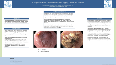
In September 2021, he was hospitalized with severe dysphagia. EGD (Figure 1) at this time showed gray, yellow frondlike lesion replacing esophageal lumen from mid to distal esophagus with mucosal friability. His dysphagia transiently responded to repeat dilations, but continued to recur and worsen.
Unfortunately, patient continued to have weight loss and dysphagia unresponsive to dilations. The patient was then started on gastric tube feedings. He was offered surgery due to the severity of his symptoms, but he declined.
Most recently, the patient underwent endoscopic mucosal resection with deeper tissue samples revealing superficial fragments of laterally spreading squamous cell carcinoma of the esophagus.
Discussion: In this case, there was a high clinical suspicion for malignancy, leading the endoscopist to obtain deeper tissue samples via endoscopic mucosal resection as superficial fragments of the esophagus only revealed parakeratosis. It is important to trust clinical judgment, especially if there is a high suspicion for malignancy.
Tylosis also has a known association with EP and often presents with areas of thickened skin plaques on palms and feet. Tylosis has a strong association with head, neck, and esophageal squamous cell carcinoma. The patient had no family history of tylosis and no cutaneous features. Despite this, a careful inspection of the entire EP area with targeted biopsies should be obtained to evaluate for dysplasia.
This case represents EP with years of no evidence of squamous cell cancer despite aggressively searching for it, which at least anecdotally suggests a precancerous potential for EP.

Disclosures:
Thomas J. Mathews, MD1, David Valadez, MD1, Madhav Desai, MD2. B0260 - A Diagnosis That Is Difficult to Swallow: Digging Deeper for Answers, ACG 2022 Annual Scientific Meeting Abstracts. Charlotte, NC: American College of Gastroenterology.
1University of Kansas Medical Center, Kansas City, KS; 2Kansas City Veterans Affairs Medical Center, Kansas City, MO
Introduction: Esophageal parakeratosis (EP) is an uncommon finding with largely unknown clinical implications and malignant potential. Here, we report a case of severe esophageal parakeratosis.
Case Description/Methods: Patient is a 69-year-old African American male with history significant alcohol and cocaine abuse, prior tobacco abuse, keloids, gastroesophageal reflux, food impaction, and progressive dysphagia for 20 years with 40-pound weight loss.
Patient’s first esophagogastroduodenoscopy (EGD) in 2000 revealed moderate esophagitis and a distal esophageal stricture with 9 mm lumen bougie dilated to 12 mm, but dysphagia persisted. Repeat EGDs between 2006 and 2016 showed whitish plaques with no evidence of fungal organisms. Given classic appearance of EOE, he was trialed on empiric fluticasone and budesonide with no improvement in dysphagia.
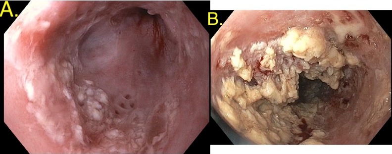
In September 2021, he was hospitalized with severe dysphagia. EGD (Figure 1) at this time showed gray, yellow frondlike lesion replacing esophageal lumen from mid to distal esophagus with mucosal friability. His dysphagia transiently responded to repeat dilations, but continued to recur and worsen.
Unfortunately, patient continued to have weight loss and dysphagia unresponsive to dilations. The patient was then started on gastric tube feedings. He was offered surgery due to the severity of his symptoms, but he declined.
Most recently, the patient underwent endoscopic mucosal resection with deeper tissue samples revealing superficial fragments of laterally spreading squamous cell carcinoma of the esophagus.
Discussion: In this case, there was a high clinical suspicion for malignancy, leading the endoscopist to obtain deeper tissue samples via endoscopic mucosal resection as superficial fragments of the esophagus only revealed parakeratosis. It is important to trust clinical judgment, especially if there is a high suspicion for malignancy.
Tylosis also has a known association with EP and often presents with areas of thickened skin plaques on palms and feet. Tylosis has a strong association with head, neck, and esophageal squamous cell carcinoma. The patient had no family history of tylosis and no cutaneous features. Despite this, a careful inspection of the entire EP area with targeted biopsies should be obtained to evaluate for dysplasia.
This case represents EP with years of no evidence of squamous cell cancer despite aggressively searching for it, which at least anecdotally suggests a precancerous potential for EP.

Figure: A. Initial EGD B. Recent EGD
Disclosures:
Thomas Mathews indicated no relevant financial relationships.
David Valadez indicated no relevant financial relationships.
Madhav Desai indicated no relevant financial relationships.
Thomas J. Mathews, MD1, David Valadez, MD1, Madhav Desai, MD2. B0260 - A Diagnosis That Is Difficult to Swallow: Digging Deeper for Answers, ACG 2022 Annual Scientific Meeting Abstracts. Charlotte, NC: American College of Gastroenterology.
