Back


Poster Session B - Monday Morning
Category: Interventional Endoscopy
B0477 - Hyperechoic Lesions on Endoscopic Ultrasound Are Not Always Benign
Monday, October 24, 2022
10:00 AM – 12:00 PM ET
Location: Crown Ballroom

Has Audio

Paul P. Hong, MD
Loyola University Medical Center
Maywood, IL
Presenting Author(s)
Paul P. Hong, MD, Abdul Haseeb, MD
Loyola University Medical Center, Maywood, IL
Introduction: Liposarcomas are rare tumors usually diagnosed in late adulthood with an uncommon incidence. Here, we present a case of a hyperechoic mass seen on the endoscopic ultrasound (EUS) with fine needle biopsies confirming a metastatic liposarcoma.
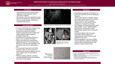
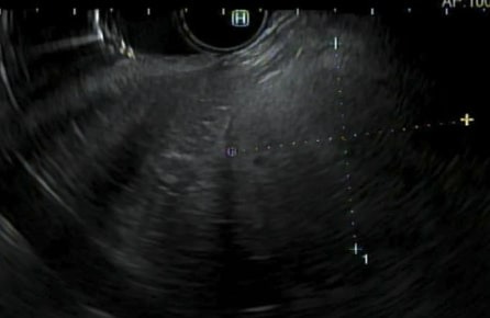
Case Description/Methods: A 70-year-old female with a distant history ( >10 years) of retroperitoneal liposarcoma with a left renal, adrenal, and colon metastasis treated with extensive surgical resection presented to a primary care clinic for health maintenance. Patient was without significant gastrointestinal symptoms or laboratory abnormalities at the visit. Patient underwent a surveillance CT of the abdomen and pelvis, where a soft tissue lesion was found in the left posterior retroperitoneal space. Patient was referred to interventional gastroenterology for EUS guided tissue biopsy. During the procedure, an oval mass was identified in the peri-splenic area at prior nephrectomy bed. The mass was hyperechoic with some isoechoic areas, 31 mm by 29 mm in maximal cross-sectional diameter, and the outer margins were not well defined (Figure 1). Fine needle biopsy using a 22 Gauge core biopsy needle was performed. Cytology analysis revealed a well-differentiated liposarcoma indicating recurrence of the disease. Patient was subsequently referred to the multidisciplinary tumor board for further evaluation and treatment.
Discussion: Interpreting echogenicity of gastrointestinal lesions with an EUS is an important part of the differential diagnosis. Lipomas are ubiquitous lesions with a classic hyperechoic appearance on EUS. Lesions with high fat or collagen content are usually hyperechogenic as a result of higher ultrasound wave scattering. Liposarcoma is a very rare soft tissue malignancy of mesenchymal origin. Four subtypes of liposarcomas exist (well-differentiated, myxoid, dedifferentiated, pleomorphic), among which a well-differentiated subtype has the largest fat content. Commonly affected sites are upper extremities, thighs, gluteal, and retroperitoneum, last of which can be detected on the EUS. Endosonographers must have a higher clinical suspicion to diagnose metastatic process while evaluating patients with history of mesenchymal tumors.

Disclosures:
Paul P. Hong, MD, Abdul Haseeb, MD. B0477 - Hyperechoic Lesions on Endoscopic Ultrasound Are Not Always Benign, ACG 2022 Annual Scientific Meeting Abstracts. Charlotte, NC: American College of Gastroenterology.
Loyola University Medical Center, Maywood, IL
Introduction: Liposarcomas are rare tumors usually diagnosed in late adulthood with an uncommon incidence. Here, we present a case of a hyperechoic mass seen on the endoscopic ultrasound (EUS) with fine needle biopsies confirming a metastatic liposarcoma.
Case Description/Methods: A 70-year-old female with a distant history ( >10 years) of retroperitoneal liposarcoma with a left renal, adrenal, and colon metastasis treated with extensive surgical resection presented to a primary care clinic for health maintenance. Patient was without significant gastrointestinal symptoms or laboratory abnormalities at the visit. Patient underwent a surveillance CT of the abdomen and pelvis, where a soft tissue lesion was found in the left posterior retroperitoneal space. Patient was referred to interventional gastroenterology for EUS guided tissue biopsy. During the procedure, an oval mass was identified in the peri-splenic area at prior nephrectomy bed. The mass was hyperechoic with some isoechoic areas, 31 mm by 29 mm in maximal cross-sectional diameter, and the outer margins were not well defined (Figure 1). Fine needle biopsy using a 22 Gauge core biopsy needle was performed. Cytology analysis revealed a well-differentiated liposarcoma indicating recurrence of the disease. Patient was subsequently referred to the multidisciplinary tumor board for further evaluation and treatment.
Discussion: Interpreting echogenicity of gastrointestinal lesions with an EUS is an important part of the differential diagnosis. Lipomas are ubiquitous lesions with a classic hyperechoic appearance on EUS. Lesions with high fat or collagen content are usually hyperechogenic as a result of higher ultrasound wave scattering. Liposarcoma is a very rare soft tissue malignancy of mesenchymal origin. Four subtypes of liposarcomas exist (well-differentiated, myxoid, dedifferentiated, pleomorphic), among which a well-differentiated subtype has the largest fat content. Commonly affected sites are upper extremities, thighs, gluteal, and retroperitoneum, last of which can be detected on the EUS. Endosonographers must have a higher clinical suspicion to diagnose metastatic process while evaluating patients with history of mesenchymal tumors.

Figure: A hyperechoic, peri-splenic mass on EUS
Disclosures:
Paul Hong indicated no relevant financial relationships.
Abdul Haseeb indicated no relevant financial relationships.
Paul P. Hong, MD, Abdul Haseeb, MD. B0477 - Hyperechoic Lesions on Endoscopic Ultrasound Are Not Always Benign, ACG 2022 Annual Scientific Meeting Abstracts. Charlotte, NC: American College of Gastroenterology.
