Back


Poster Session D - Tuesday Morning
Category: IBD
D0419 - Eye-BD— A Rare Case of Orbital Myositis as an Extra-intestinal Manifestation of Crohn’s Disease
Tuesday, October 25, 2022
10:00 AM – 12:00 PM ET
Location: Crown Ballroom

Has Audio
- IR
Ian Robertson, MD
Walter Reed National Military Medical Center
Bethesda, MD
Presenting Author(s)
Kevin Pak, MD1, Joseph Cheatham, MD1, Ian Robertson, MD2, Rashad C. Wilkerson, DO1, Morgan Harvey, MD1, Katie Topping, MD1
1Naval Medical Center San Diego, San Diego, CA; 2Walter Reed National Military Medical Center, Bethesda, MD
Introduction: Crohn’s Disease (CD) is often associated with a diverse array of extra-intestinal manifestations (EIMs). The estimated prevalence of EIMs in patients with CD is 25-70%. The estimated prevalence of ocular manifestations in patients with CD is 3.5-6.8%. Orbital myositis (OM), characterized by inflammation of the extraocular muscles, is a rare ocular EIM associated with CD.
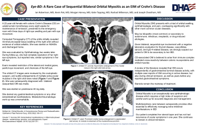
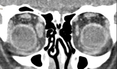
Case Description/Methods: A 22-year-old female with CD on ustekinumab for maintenance therapy in remission presented with three days of right eye swelling and pain with extraocular movements. CT of the orbits reported minimal pre-septal tissue swelling, without evidence of orbital cellulitis. She was started on NSAIDs. A week later, the patient presented to the Ophthalmology clinic with resolution of her right eye symptoms but with new, similar symptoms in her left eye, including diplopia, restriction of lateral and medial gaze, and chemosis of the lateral conjunctiva. She denied symptoms concerning for a Crohn’s flare or other EIMs. The initial CT of the orbits was reviewed, and an addendum was made, reporting subtle enlargements of multiple extra-ocular muscles in the right orbit. She was diagnosed with bilateral OM. Infectious, endocrine, and autoimmune work-up was performed which were unrevealing and her OM was likely an EIM of her CD. She was started on prednisone 40 mg daily, which resulted in complete resolution of her symptoms at one-week follow-up. She was kept on ustekinumab maintenance therapy as she remained in clinical remission without evidence of other EIMs and completed her steroid taper without further complication. The patient was informed of the high recurrence rate of CD-associated OM and close follow-up was arranged.
Discussion: Overall, our patient’s OM was a likely EIM of her CD, as further laboratory work-up for commonly implicated etiologies to include thyroid disease, vasculitides, sarcoidosis, and IgG4-related disease was all negative. This case illustrates a rare EIM of CD in a patient believed to be in clinical remission, emphasizing the point that certain EIMs may occur independently of gastrointestinal luminal disease activity. Given that a high index of suspicion was necessary to reach the diagnosis given the subtle exam and imaging findings, this case also reiterates the importance of timely diagnosis and early management to minimize morbidity risk, which can be best accomplished through multidisciplinary coordination among subspecialty providers.

Disclosures:
Kevin Pak, MD1, Joseph Cheatham, MD1, Ian Robertson, MD2, Rashad C. Wilkerson, DO1, Morgan Harvey, MD1, Katie Topping, MD1. D0419 - Eye-BD— A Rare Case of Orbital Myositis as an Extra-intestinal Manifestation of Crohn’s Disease, ACG 2022 Annual Scientific Meeting Abstracts. Charlotte, NC: American College of Gastroenterology.
1Naval Medical Center San Diego, San Diego, CA; 2Walter Reed National Military Medical Center, Bethesda, MD
Introduction: Crohn’s Disease (CD) is often associated with a diverse array of extra-intestinal manifestations (EIMs). The estimated prevalence of EIMs in patients with CD is 25-70%. The estimated prevalence of ocular manifestations in patients with CD is 3.5-6.8%. Orbital myositis (OM), characterized by inflammation of the extraocular muscles, is a rare ocular EIM associated with CD.
Case Description/Methods: A 22-year-old female with CD on ustekinumab for maintenance therapy in remission presented with three days of right eye swelling and pain with extraocular movements. CT of the orbits reported minimal pre-septal tissue swelling, without evidence of orbital cellulitis. She was started on NSAIDs. A week later, the patient presented to the Ophthalmology clinic with resolution of her right eye symptoms but with new, similar symptoms in her left eye, including diplopia, restriction of lateral and medial gaze, and chemosis of the lateral conjunctiva. She denied symptoms concerning for a Crohn’s flare or other EIMs. The initial CT of the orbits was reviewed, and an addendum was made, reporting subtle enlargements of multiple extra-ocular muscles in the right orbit. She was diagnosed with bilateral OM. Infectious, endocrine, and autoimmune work-up was performed which were unrevealing and her OM was likely an EIM of her CD. She was started on prednisone 40 mg daily, which resulted in complete resolution of her symptoms at one-week follow-up. She was kept on ustekinumab maintenance therapy as she remained in clinical remission without evidence of other EIMs and completed her steroid taper without further complication. The patient was informed of the high recurrence rate of CD-associated OM and close follow-up was arranged.
Discussion: Overall, our patient’s OM was a likely EIM of her CD, as further laboratory work-up for commonly implicated etiologies to include thyroid disease, vasculitides, sarcoidosis, and IgG4-related disease was all negative. This case illustrates a rare EIM of CD in a patient believed to be in clinical remission, emphasizing the point that certain EIMs may occur independently of gastrointestinal luminal disease activity. Given that a high index of suspicion was necessary to reach the diagnosis given the subtle exam and imaging findings, this case also reiterates the importance of timely diagnosis and early management to minimize morbidity risk, which can be best accomplished through multidisciplinary coordination among subspecialty providers.

Figure: Orbital CT, Coronal- Asymmetric enlargement of the medial rectus in the RT orbit.
Disclosures:
Kevin Pak indicated no relevant financial relationships.
Joseph Cheatham indicated no relevant financial relationships.
Ian Robertson indicated no relevant financial relationships.
Rashad Wilkerson indicated no relevant financial relationships.
Morgan Harvey indicated no relevant financial relationships.
Katie Topping indicated no relevant financial relationships.
Kevin Pak, MD1, Joseph Cheatham, MD1, Ian Robertson, MD2, Rashad C. Wilkerson, DO1, Morgan Harvey, MD1, Katie Topping, MD1. D0419 - Eye-BD— A Rare Case of Orbital Myositis as an Extra-intestinal Manifestation of Crohn’s Disease, ACG 2022 Annual Scientific Meeting Abstracts. Charlotte, NC: American College of Gastroenterology.
