Back


Poster Session D - Tuesday Morning
Category: GI Bleeding
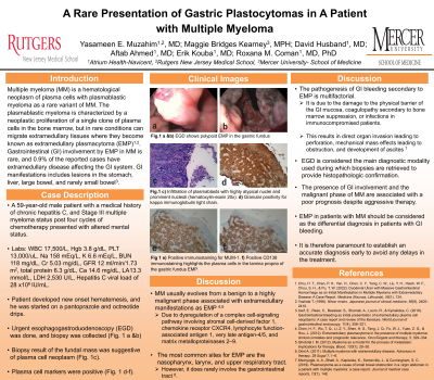
D0313 - A Rare Presentation of Gastric Plastocytomas in a Patient With Multiple Myeloma
Tuesday, October 25, 2022
10:00 AM – 12:00 PM ET
Location: Crown Ballroom

Has Audio

Yasameen E. Muzahim, MD
Atrium Health Navicent
Presenting Author(s)
Yasameen E. Muzahim, MD1, Maggie B. Kearney, MPH2, David Husband, MD1, Aftab Ahmed, MD1, Erik Kouba, MD1, Roxana M. Coman, MD, PhD1
1Atrium Health Navicent, Macon, GA; 2Mercer University, School of Medicine, Macon, GA
Introduction: Multiple myeloma is an uncommon hematological neoplasm. This malignancy involves unchecked plasma cell growth in the bone marrow, but in a subset of often more advanced cases, these cells may migrate to soft tissues causing extramedullary plasmacytoma. This is unusual, and, in the literature, few cases of extramedullary plasmacytomas involve the gastrointestinal tract. Within this system, the more common locations are the stomach, the liver, the large bowel, and more rarely, the small bowel.
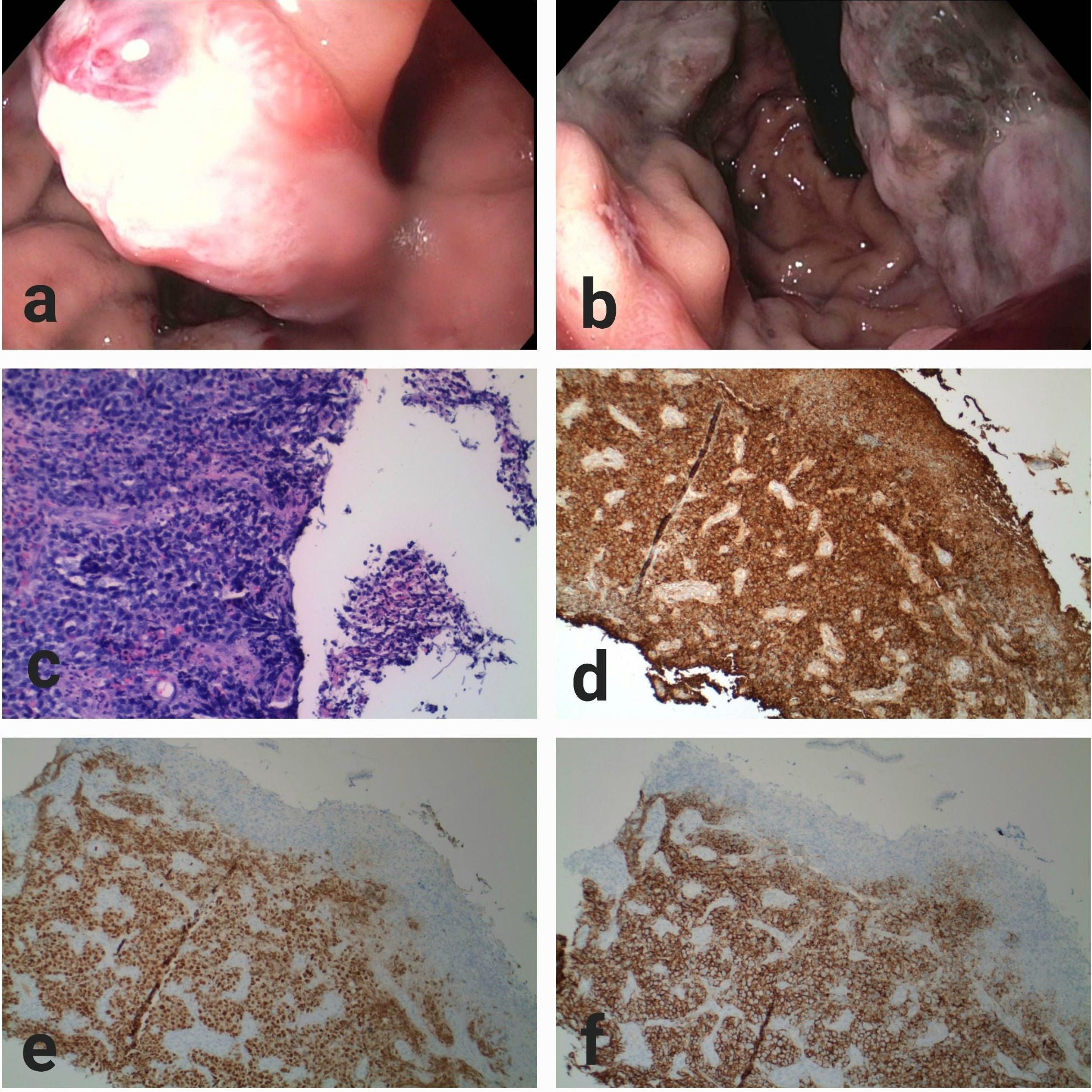
Case Description/Methods: A 59-year-old male with a medical history of chronic hepatitis C, and Stage III multiple myeloma status post four cycles of chemotherapy presented with altered mental status. Upon evaluation, he had significant electrolyte abnormalities, remarkable anemia, severe renal and liver dysfunction, and a positive hemoccult test. Our patient received intravenous fluid, and multiple blood products. He was then admitted to the intensive care unit for a high level of care. On the second day of admission, he developed new onset hematemesis, and the gastroenterology team was consulted. The patient was started on a pantoprazole drip. Urgent esophagogastroduodenoscopy showed several polypoid appearing masses with overlying ulceration in the gastric fundus (Fig. 1 a &b), and tissue biopsies were obtained. Biopsy result of the polypoid fundal mass showed a large cell neoplasm with plasmocytic differentiation. Gastric tissue sections showed an ulcerated lesion with submucosal, tightly packed atypical large cells with multinucleation, prominent nucleoli and relatively identifiable mitoses (Fig. 1c). A panel of immunohistochemical stains with plasma cell markers were positive for kappa, Multiple Myloma-1 nuclear protein, and CD138 (Fig. 1 d-f). The overall findings were compatible with gastric involvement by a plasma cell neoplasm.
Discussion: This case highlights a complicated, yet a uniquely rare presentation of extramedullary plasmacytoma in a patient diagnosed with multiple myeloma. Although the gastrointestinal tract is rarely involved in patients with multiple myeloma, it should be considered as the differential diagnosis in patients with gastrointestinal bleeding especially with a known concurrent systemic hematological malignancy. The presence of gastrointestinal involvement and the malignant phase of multiple myeloma are associated with a poor prognosis despite aggressive therapy, and it is therefore paramount to establish an accurate diagnosis early to avoid any delays in treatment.

Disclosures:
Yasameen E. Muzahim, MD1, Maggie B. Kearney, MPH2, David Husband, MD1, Aftab Ahmed, MD1, Erik Kouba, MD1, Roxana M. Coman, MD, PhD1. D0313 - A Rare Presentation of Gastric Plastocytomas in a Patient With Multiple Myeloma, ACG 2022 Annual Scientific Meeting Abstracts. Charlotte, NC: American College of Gastroenterology.
1Atrium Health Navicent, Macon, GA; 2Mercer University, School of Medicine, Macon, GA
Introduction: Multiple myeloma is an uncommon hematological neoplasm. This malignancy involves unchecked plasma cell growth in the bone marrow, but in a subset of often more advanced cases, these cells may migrate to soft tissues causing extramedullary plasmacytoma. This is unusual, and, in the literature, few cases of extramedullary plasmacytomas involve the gastrointestinal tract. Within this system, the more common locations are the stomach, the liver, the large bowel, and more rarely, the small bowel.
Case Description/Methods: A 59-year-old male with a medical history of chronic hepatitis C, and Stage III multiple myeloma status post four cycles of chemotherapy presented with altered mental status. Upon evaluation, he had significant electrolyte abnormalities, remarkable anemia, severe renal and liver dysfunction, and a positive hemoccult test. Our patient received intravenous fluid, and multiple blood products. He was then admitted to the intensive care unit for a high level of care. On the second day of admission, he developed new onset hematemesis, and the gastroenterology team was consulted. The patient was started on a pantoprazole drip. Urgent esophagogastroduodenoscopy showed several polypoid appearing masses with overlying ulceration in the gastric fundus (Fig. 1 a &b), and tissue biopsies were obtained. Biopsy result of the polypoid fundal mass showed a large cell neoplasm with plasmocytic differentiation. Gastric tissue sections showed an ulcerated lesion with submucosal, tightly packed atypical large cells with multinucleation, prominent nucleoli and relatively identifiable mitoses (Fig. 1c). A panel of immunohistochemical stains with plasma cell markers were positive for kappa, Multiple Myloma-1 nuclear protein, and CD138 (Fig. 1 d-f). The overall findings were compatible with gastric involvement by a plasma cell neoplasm.
Discussion: This case highlights a complicated, yet a uniquely rare presentation of extramedullary plasmacytoma in a patient diagnosed with multiple myeloma. Although the gastrointestinal tract is rarely involved in patients with multiple myeloma, it should be considered as the differential diagnosis in patients with gastrointestinal bleeding especially with a known concurrent systemic hematological malignancy. The presence of gastrointestinal involvement and the malignant phase of multiple myeloma are associated with a poor prognosis despite aggressive therapy, and it is therefore paramount to establish an accurate diagnosis early to avoid any delays in treatment.

Figure: Fig. 1 a&b) EGD shows polypoid EMP in the gastric fundus. c) Infiltration of plasmablasts with highly atypical nuclei and prominent nucleoli (hematoxylin-eosin 20x). d) Granular positivity for kappa immunoglobulin light chain. e) Positive immunostaining for MUM-1. f) Positive CD138 immunostaining highlights the plasma cells in the lamina propria of the gastric fundus EMP.
Disclosures:
Yasameen Muzahim indicated no relevant financial relationships.
Maggie Kearney indicated no relevant financial relationships.
David Husband indicated no relevant financial relationships.
Aftab Ahmed indicated no relevant financial relationships.
Erik Kouba indicated no relevant financial relationships.
Roxana Coman indicated no relevant financial relationships.
Yasameen E. Muzahim, MD1, Maggie B. Kearney, MPH2, David Husband, MD1, Aftab Ahmed, MD1, Erik Kouba, MD1, Roxana M. Coman, MD, PhD1. D0313 - A Rare Presentation of Gastric Plastocytomas in a Patient With Multiple Myeloma, ACG 2022 Annual Scientific Meeting Abstracts. Charlotte, NC: American College of Gastroenterology.
