Back


Poster Session C - Monday Afternoon
Category: Interventional Endoscopy
C0484 - A Case of a Completely Migrated Gastric Toothpick Caught by EUS
Monday, October 24, 2022
3:00 PM – 5:00 PM ET
Location: Crown Ballroom

Has Audio

Andrew Mims, MD
The University of Tennessee Health Science Center
Chattanooga, TN
Presenting Author(s)
Andrew Mims, MD1, Aparna Shreenath, MD1, George Philips, MD1, Peter Deleeuw, DO1, Steven Kessler, DO2, Sharif Murphy, MD1
1University of Tennessee Health Science Center, Chattanooga, TN; 2The University of Tennessee Health Science Center, Chattanooga, TN
Introduction: Foreign body ingestion is a common cause for medical emergencies. Once they reach the stomach, roughly 80% of foreign bodies pass uneventfully through the GI tract. Sharp objects, such as toothpicks, are more likely to cause injury throughout the GI tract. Toothpick ingestion is associated with high rates of mortality and morbidity, with estimated 79% of ingestions leading to gut perforation. Initial diagnosis can be difficult, as ingestion has been found to mimic other conditions, such as renal colic. Endoscopy is warranted in roughly 20% of cases, with about 1% requiring surgical intervention.
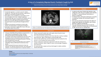
Case Description/Methods: A 63-year-old male with a history of HTN and DM presented with a 1-week history of altered mental status, fevers, nausea, vomiting, and melena. CT revealed a linear density extending from the lumen of the distal gastric antrum to the posterior wall of the stomach traversing the pancreas in the expected location of the SMV. Follow up EGD and MRI failed to reveal foreign body. He subsequently developed actinomyces bacteremia, was treated with antibiotics along with dental extraction, and was discharged home. The patient later returned with septic shock, found to have recurrent Actinomyces bacteremia. CT re-demonstrated suspicious thin linear density as well as multiple liver abscesses. Again, EGD failed to reveal foreign body. This time, an EUS was performed, and the foreign body was localized to the antrum, 2.4 mm below the luminal surface. Endoscopic mucosal resection was attempted but was unsuccessful. The patient then underwent an exploratory laparotomy with the retrieval of a wooden toothpick. He discharged in stable condition.
Discussion: In this case, original CT scans revealed a possible foreign body, the object being localized as expected near the SMV. Given original negative EGD and MRI, it was felt that CT scan findings represented vasculature. When patient returned in septic shock with recurrent actinomyces bacteremia, a second EGD was performed and was negative. A decision was made at that time to investigate with EUS as a diagnostic tool, which ultimately revealed the location of the foreign body. What makes our case particularly unique is the degree of foreign body migration into the gastric wall, causing difficulty in diagnosis through EGD. This case highlights EUS as an important diagnostic modality in cases of foreign body ingestion.
Disclosures:
Andrew Mims, MD1, Aparna Shreenath, MD1, George Philips, MD1, Peter Deleeuw, DO1, Steven Kessler, DO2, Sharif Murphy, MD1. C0484 - A Case of a Completely Migrated Gastric Toothpick Caught by EUS, ACG 2022 Annual Scientific Meeting Abstracts. Charlotte, NC: American College of Gastroenterology.
1University of Tennessee Health Science Center, Chattanooga, TN; 2The University of Tennessee Health Science Center, Chattanooga, TN
Introduction: Foreign body ingestion is a common cause for medical emergencies. Once they reach the stomach, roughly 80% of foreign bodies pass uneventfully through the GI tract. Sharp objects, such as toothpicks, are more likely to cause injury throughout the GI tract. Toothpick ingestion is associated with high rates of mortality and morbidity, with estimated 79% of ingestions leading to gut perforation. Initial diagnosis can be difficult, as ingestion has been found to mimic other conditions, such as renal colic. Endoscopy is warranted in roughly 20% of cases, with about 1% requiring surgical intervention.
Case Description/Methods: A 63-year-old male with a history of HTN and DM presented with a 1-week history of altered mental status, fevers, nausea, vomiting, and melena. CT revealed a linear density extending from the lumen of the distal gastric antrum to the posterior wall of the stomach traversing the pancreas in the expected location of the SMV. Follow up EGD and MRI failed to reveal foreign body. He subsequently developed actinomyces bacteremia, was treated with antibiotics along with dental extraction, and was discharged home. The patient later returned with septic shock, found to have recurrent Actinomyces bacteremia. CT re-demonstrated suspicious thin linear density as well as multiple liver abscesses. Again, EGD failed to reveal foreign body. This time, an EUS was performed, and the foreign body was localized to the antrum, 2.4 mm below the luminal surface. Endoscopic mucosal resection was attempted but was unsuccessful. The patient then underwent an exploratory laparotomy with the retrieval of a wooden toothpick. He discharged in stable condition.
Discussion: In this case, original CT scans revealed a possible foreign body, the object being localized as expected near the SMV. Given original negative EGD and MRI, it was felt that CT scan findings represented vasculature. When patient returned in septic shock with recurrent actinomyces bacteremia, a second EGD was performed and was negative. A decision was made at that time to investigate with EUS as a diagnostic tool, which ultimately revealed the location of the foreign body. What makes our case particularly unique is the degree of foreign body migration into the gastric wall, causing difficulty in diagnosis through EGD. This case highlights EUS as an important diagnostic modality in cases of foreign body ingestion.
Disclosures:
Andrew Mims indicated no relevant financial relationships.
Aparna Shreenath indicated no relevant financial relationships.
George Philips indicated no relevant financial relationships.
Peter Deleeuw indicated no relevant financial relationships.
Steven Kessler indicated no relevant financial relationships.
Sharif Murphy indicated no relevant financial relationships.
Andrew Mims, MD1, Aparna Shreenath, MD1, George Philips, MD1, Peter Deleeuw, DO1, Steven Kessler, DO2, Sharif Murphy, MD1. C0484 - A Case of a Completely Migrated Gastric Toothpick Caught by EUS, ACG 2022 Annual Scientific Meeting Abstracts. Charlotte, NC: American College of Gastroenterology.
