Back


Poster Session C - Monday Afternoon
Category: Interventional Endoscopy
C0466 - Endoscopic Ultrasound-Guided Biopsy of Non-Functioning Retropancreatic Celiac Paraganglioma Masquerading as Malignant Lymph Node
Monday, October 24, 2022
3:00 PM – 5:00 PM ET
Location: Crown Ballroom

Has Audio

Monica Dzwonkowski, DO
Geisinger Medical Center
Danville, PA
Presenting Author(s)
Monica Dzwonkowski, DO1, Sunny Patel, DO1, Yazan Hasan, MD1, Harshit S. Khara, MD, FACG2
1Geisinger Medical Center, Danville, PA; 2Geisinger Health System, Danville, PA
Introduction: Paragangliomas are rare, extra-adrenal neuroendocrine tumors derived from chromaffin cells. Pheochromocytomas are in the adrenal medulla, whereas paragangliomas are extra-adrenal and can be found anywhere in the body. We present a unique case of a woman presenting with abdominal pain, found to have a micro-lobulated mass at the bifurcation of the celiac axis on imaging, suspected to be metastatic lymphadenopathy. She was referred for endoscopic ultrasound (EUS) guided fine-needle biopsy which revealed the mass to be a paraganglioma.
Case Description/Methods: A 59-year-old female with a history of chronic kidney disease and tobacco use presented to her primary care physician’s office with complaint of abdominal fullness, diffuse abdominal pain, and urinary frequency. She underwent CT imaging of her abdomen and pelvis which revealed a 2.8 cm mass with micro-lobulated margins located at the bifurcation of the celiac axis (Fig 1A, 1B). A follow up PET scan revealed increased uptake by the mass with a standardized uptake value of 6.7 (Fig 1C). Due to the delicate location of the lesion, the patient was referred to gastroenterology for biopsy of the mass. She underwent EUS-guided fine-needle biopsy (FNB) using a 25G FNB needle which revealed two hypoechoic, triangular masses (20mm and 10mm) in the celiac region thought to be lymph nodes (Fig 1D, 1E). Pathology was consistent with paraganglioma. Lab workup revealed a urinary free cortisol of 56mcg/24hr (ref: 4.0 to 50.0 mcg/24hr) and normal urine catecholamines. She opted to see an endocrine surgical specialist and is undergoing surveillance with potential future surgical resection.
Discussion: Paragangliomas are a rare phenomenon and may be misdiagnosed as patients can have nonspecific findings and the lesions vary in location. Most masses are benign but 15-20% may be malignant, thus clinicians should maintain a high index of suspicion for paraganglioma in patients with unexplained abdominal masses. Imaging and biopsy are the mainstays of diagnosis. Endoscopic ultrasound is a useful modality in obtaining sampling via FNB, especially from difficult to reach lesions as was the case for our patient.

Disclosures:
Monica Dzwonkowski, DO1, Sunny Patel, DO1, Yazan Hasan, MD1, Harshit S. Khara, MD, FACG2. C0466 - Endoscopic Ultrasound-Guided Biopsy of Non-Functioning Retropancreatic Celiac Paraganglioma Masquerading as Malignant Lymph Node, ACG 2022 Annual Scientific Meeting Abstracts. Charlotte, NC: American College of Gastroenterology.
1Geisinger Medical Center, Danville, PA; 2Geisinger Health System, Danville, PA
Introduction: Paragangliomas are rare, extra-adrenal neuroendocrine tumors derived from chromaffin cells. Pheochromocytomas are in the adrenal medulla, whereas paragangliomas are extra-adrenal and can be found anywhere in the body. We present a unique case of a woman presenting with abdominal pain, found to have a micro-lobulated mass at the bifurcation of the celiac axis on imaging, suspected to be metastatic lymphadenopathy. She was referred for endoscopic ultrasound (EUS) guided fine-needle biopsy which revealed the mass to be a paraganglioma.
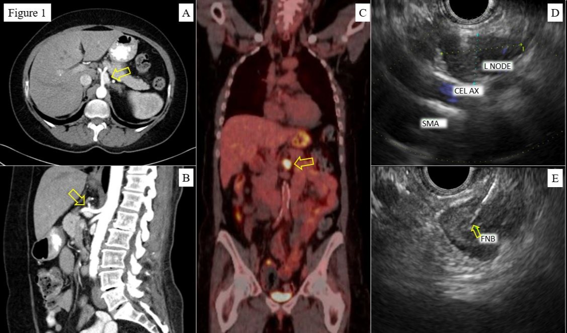
Case Description/Methods: A 59-year-old female with a history of chronic kidney disease and tobacco use presented to her primary care physician’s office with complaint of abdominal fullness, diffuse abdominal pain, and urinary frequency. She underwent CT imaging of her abdomen and pelvis which revealed a 2.8 cm mass with micro-lobulated margins located at the bifurcation of the celiac axis (Fig 1A, 1B). A follow up PET scan revealed increased uptake by the mass with a standardized uptake value of 6.7 (Fig 1C). Due to the delicate location of the lesion, the patient was referred to gastroenterology for biopsy of the mass. She underwent EUS-guided fine-needle biopsy (FNB) using a 25G FNB needle which revealed two hypoechoic, triangular masses (20mm and 10mm) in the celiac region thought to be lymph nodes (Fig 1D, 1E). Pathology was consistent with paraganglioma. Lab workup revealed a urinary free cortisol of 56mcg/24hr (ref: 4.0 to 50.0 mcg/24hr) and normal urine catecholamines. She opted to see an endocrine surgical specialist and is undergoing surveillance with potential future surgical resection.
Discussion: Paragangliomas are a rare phenomenon and may be misdiagnosed as patients can have nonspecific findings and the lesions vary in location. Most masses are benign but 15-20% may be malignant, thus clinicians should maintain a high index of suspicion for paraganglioma in patients with unexplained abdominal masses. Imaging and biopsy are the mainstays of diagnosis. Endoscopic ultrasound is a useful modality in obtaining sampling via FNB, especially from difficult to reach lesions as was the case for our patient.

Figure: Figure 1A,B: Computed tomography images showing abdominal mass near celiac bifurcation, transverse (A) and sagittal (B) planes
Figure 1C: Positron emission tomography scan revealing enhancement of abdominal mass, coronal view
Figure 1D: Endoscopic ultrasound image of mass suspected to be lymph node near celiac axis
Figure 1E: Endoscopic ultrasound guided fine-needle biopsy of mass
Figure 1C: Positron emission tomography scan revealing enhancement of abdominal mass, coronal view
Figure 1D: Endoscopic ultrasound image of mass suspected to be lymph node near celiac axis
Figure 1E: Endoscopic ultrasound guided fine-needle biopsy of mass
Disclosures:
Monica Dzwonkowski indicated no relevant financial relationships.
Sunny Patel indicated no relevant financial relationships.
Yazan Hasan indicated no relevant financial relationships.
Harshit Khara indicated no relevant financial relationships.
Monica Dzwonkowski, DO1, Sunny Patel, DO1, Yazan Hasan, MD1, Harshit S. Khara, MD, FACG2. C0466 - Endoscopic Ultrasound-Guided Biopsy of Non-Functioning Retropancreatic Celiac Paraganglioma Masquerading as Malignant Lymph Node, ACG 2022 Annual Scientific Meeting Abstracts. Charlotte, NC: American College of Gastroenterology.
