Back


Poster Session D - Tuesday Morning
Category: Interventional Endoscopy
D0465 - Duodenal Stent Fixation Using Through-the-Scope Helix Tack and Suturing Device
Tuesday, October 25, 2022
10:00 AM – 12:00 PM ET
Location: Crown Ballroom

Has Audio
.jpg)
Natalie Wilson, MD
University of Minnesota
Minneapolis, MN
Presenting Author(s)
Natalie Wilson, MD1, Nicholas McDonald, MD1, Bryant Megna, MD1, Mohamed Abdallah, MD2, Mohammad Bilal, MD3
1University of Minnesota, Minneapolis, MN; 2University of Minnesota Medical Center, Minneapolis, MN; 3University of Minnesota, Minneapolis VA Medical Center, Minneapolis, MN
Introduction: Malignant gastroduodenal strictures are a leading cause of gastric outlet obstruction and are often managed with endoscopic stent placement. One of the main limitations of duodenal self-expanding metal stents (SEMS) is the risk of migration. Multiple techniques have been used to prevent stent migration, including stent fixation using through-the-scope (TTS) clips, over-the-scope stent fixation devices, and endoscopic suturing. TTS suturing using the helix tack and suturing device is a novel suturing method that is generally used for defect closure, though it has been rapidly gaining popularity for alternate uses. Here, we report its use as a stent fixation method in a patient with gastric outlet obstruction due to a malignant duodenal stricture.
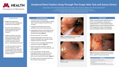
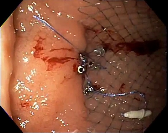
Case Description/Methods: A 73-year-old male with pancreatic adenocarcinoma on neoadjuvant chemotherapy presented with 3 weeks of vomiting and abdominal distension. Imaging showed pancreatic adenocarcinoma with duodenal obstruction. Esophagogastroduodenoscopy (EGD) was performed and showed duodenal stenosis from tumor infiltration at the duodenal sweep. The adult upper endoscope was able to traverse the stenosis with significant resistance and the stenosis measured 2 cm in length. The patient was borderline resectable, and per institutional protocol, endoscopic ultrasound-guided gastrojejunostomy is only performed for patients who are not surgical candidates, therefore, decision was made to proceed with duodenal stent placement. A 25 mm in diameter and 10 cm in length uncovered SEMS was placed across the stenosis. Given that the upper endoscope was able to traverse the stenosis, decision was made to fixate the stent to reduce migration prior to stent expansion and tissue ingrowth to keep the stent in place. The TTS suturing device was used and the stent was fixated with four tacks placed in stent-mucosa-mucosa-stent fashion (Figure 1). A cinch was placed in the end. The patient tolerated the procedure well and no adverse events were reported within the first 4 weeks of the procedure.
Discussion: Duodenal SEMS are widely used in the setting of malignant gastroduodenal obstruction. While uncovered stents typically carry a lower risk of migration compared to covered stents, in this case, the tissue apposition was less than desired and thus the TTS suturing device was used for stent fixation. In this report, we demonstrate that the TTS suturing device may be a safe and effective technique for duodenal stent fixation.

Disclosures:
Natalie Wilson, MD1, Nicholas McDonald, MD1, Bryant Megna, MD1, Mohamed Abdallah, MD2, Mohammad Bilal, MD3. D0465 - Duodenal Stent Fixation Using Through-the-Scope Helix Tack and Suturing Device, ACG 2022 Annual Scientific Meeting Abstracts. Charlotte, NC: American College of Gastroenterology.
1University of Minnesota, Minneapolis, MN; 2University of Minnesota Medical Center, Minneapolis, MN; 3University of Minnesota, Minneapolis VA Medical Center, Minneapolis, MN
Introduction: Malignant gastroduodenal strictures are a leading cause of gastric outlet obstruction and are often managed with endoscopic stent placement. One of the main limitations of duodenal self-expanding metal stents (SEMS) is the risk of migration. Multiple techniques have been used to prevent stent migration, including stent fixation using through-the-scope (TTS) clips, over-the-scope stent fixation devices, and endoscopic suturing. TTS suturing using the helix tack and suturing device is a novel suturing method that is generally used for defect closure, though it has been rapidly gaining popularity for alternate uses. Here, we report its use as a stent fixation method in a patient with gastric outlet obstruction due to a malignant duodenal stricture.
Case Description/Methods: A 73-year-old male with pancreatic adenocarcinoma on neoadjuvant chemotherapy presented with 3 weeks of vomiting and abdominal distension. Imaging showed pancreatic adenocarcinoma with duodenal obstruction. Esophagogastroduodenoscopy (EGD) was performed and showed duodenal stenosis from tumor infiltration at the duodenal sweep. The adult upper endoscope was able to traverse the stenosis with significant resistance and the stenosis measured 2 cm in length. The patient was borderline resectable, and per institutional protocol, endoscopic ultrasound-guided gastrojejunostomy is only performed for patients who are not surgical candidates, therefore, decision was made to proceed with duodenal stent placement. A 25 mm in diameter and 10 cm in length uncovered SEMS was placed across the stenosis. Given that the upper endoscope was able to traverse the stenosis, decision was made to fixate the stent to reduce migration prior to stent expansion and tissue ingrowth to keep the stent in place. The TTS suturing device was used and the stent was fixated with four tacks placed in stent-mucosa-mucosa-stent fashion (Figure 1). A cinch was placed in the end. The patient tolerated the procedure well and no adverse events were reported within the first 4 weeks of the procedure.
Discussion: Duodenal SEMS are widely used in the setting of malignant gastroduodenal obstruction. While uncovered stents typically carry a lower risk of migration compared to covered stents, in this case, the tissue apposition was less than desired and thus the TTS suturing device was used for stent fixation. In this report, we demonstrate that the TTS suturing device may be a safe and effective technique for duodenal stent fixation.

Figure: Figure 1. The duodenal stent has been fixated with four tacks placed in a stent-mucosa-mucosa-stent fashion
Disclosures:
Natalie Wilson indicated no relevant financial relationships.
Nicholas McDonald indicated no relevant financial relationships.
Bryant Megna indicated no relevant financial relationships.
Mohamed Abdallah indicated no relevant financial relationships.
Mohammad Bilal indicated no relevant financial relationships.
Natalie Wilson, MD1, Nicholas McDonald, MD1, Bryant Megna, MD1, Mohamed Abdallah, MD2, Mohammad Bilal, MD3. D0465 - Duodenal Stent Fixation Using Through-the-Scope Helix Tack and Suturing Device, ACG 2022 Annual Scientific Meeting Abstracts. Charlotte, NC: American College of Gastroenterology.
