Back


Poster Session C - Monday Afternoon
Category: Stomach
C0735 - Sarcina Ventriculi Infection: A Rare Cause of Gastric Outlet Obstruction
Monday, October 24, 2022
3:00 PM – 5:00 PM ET
Location: Crown Ballroom

Has Audio

Vijayvardhan Kamalumpundi, BS
University of Iowa Carver College of Medicine
Iowa City, IA
Presenting Author(s)
Vijayvardhan Kamalumpundi, BS1, Katelin M. Durham, MD2, Steven Polyak, MD2, Joseph Laakman, MD2, Aditi Reddy, MD3, Xiaocen Zhang, MD4
1University of Iowa Carver College of Medicine, Iowa City, IA; 2University of Iowa Hospitals and Clinics, Iowa City, IA; 3UIHC, Iowa City, IA; 4University of Iowa Hospitals & Clinics, Iowa City, IA
Introduction: Sarcina ventriculi is an anaerobic, gram-positive coccus that grows in acidic environments including the stomach. Case reports have implicated its role in causing gastric ulcers, emphysematous gastritis, and gastric perforation. There has been only one case report in the literature on S. ventriculi causing gastric mass lesion to our knowledge [1]. Here we report a rare case of S. ventriculi infection causing a pyloric mass leading to gastric outlet obstruction (GOO).
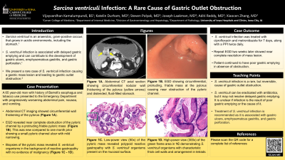
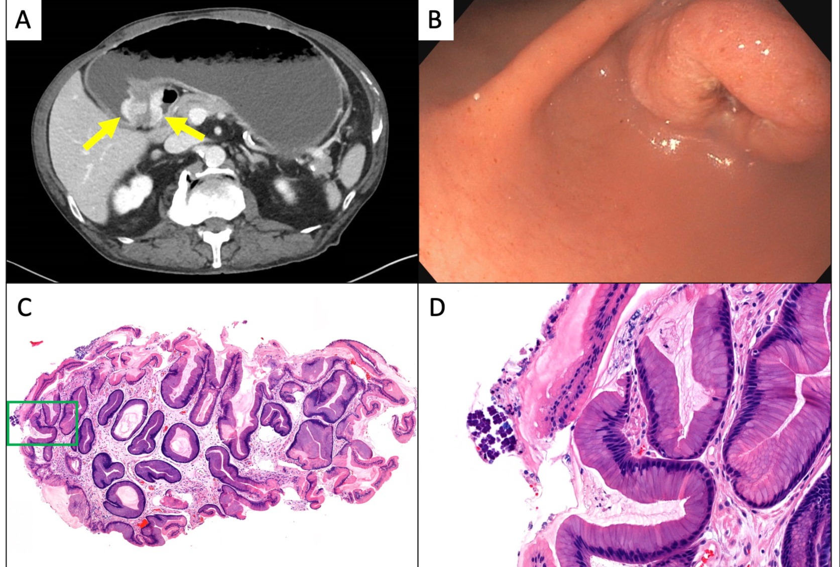
Case Description/Methods: A 65-year-old male with a past medical history of Barrett’s esophagus and tobacco use presented to the emergency department with progressive worsening of abdominal pain, nausea, and vomiting. Esophagogastroduodenoscopy (EGD) performed locally showed a small pyloric channel ulcer with traversable pyloric narrowing. Gastric biopsies showed no significant pathology and were negative for H. pylori. Abdominal computed tomography showed circumferential nodular wall thickening of the pylorus and a dilated, fluid-filled stomach consistent with GOO (Figure 1A). No pneumatosis or perforation of the stomach was noted. EGD was repeated one month later at our institution due to worsening symptoms and revealed near complete obstruction of the pyloric channel by a protruding friable pyloric mass (Figure 1B). Biopsies of the mass revealed S.ventriculi organisms in the background of reactive gastropathy, with no evidence of malignancy (Figure 1C, 1D). A nasojejunal feeding tube was placed and the patient was treated with ciprofloxacin and metronidazole for 7 days along with pantoprazole twice daily. Repeat EGD performed two weeks later showed near complete resolution of the mass lesion.
Discussion: S. ventriculi infections are associated with delayed gastric emptying [1]. It is unclear if the infection is a result of the poor gastric emptying or the cause of it. In our case the patient presented with an obstructing pyloric mass due to reactive gastropathy in the setting of S.ventriculi infection, and repeat EGD after treatment with antibiotics and a proton pump inhibitor showed near complete healing of the mass and ulcer but persistent poor gastric emptying in the absence of obstruction. We report this case to expand on the paucity of literature regarding S. ventriculi gastrointestinal infections and to raise awareness of it presenting as a gastric mass lesion.
Ref: [1] Tartaglia et al. (2022). Sarcina ventriculi infection: a rare but fearsome event. A Systematic Review of the Literature. Int J of Infect Dis. 115, 48-61.

Disclosures:
Vijayvardhan Kamalumpundi, BS1, Katelin M. Durham, MD2, Steven Polyak, MD2, Joseph Laakman, MD2, Aditi Reddy, MD3, Xiaocen Zhang, MD4. C0735 - Sarcina Ventriculi Infection: A Rare Cause of Gastric Outlet Obstruction, ACG 2022 Annual Scientific Meeting Abstracts. Charlotte, NC: American College of Gastroenterology.
1University of Iowa Carver College of Medicine, Iowa City, IA; 2University of Iowa Hospitals and Clinics, Iowa City, IA; 3UIHC, Iowa City, IA; 4University of Iowa Hospitals & Clinics, Iowa City, IA
Introduction: Sarcina ventriculi is an anaerobic, gram-positive coccus that grows in acidic environments including the stomach. Case reports have implicated its role in causing gastric ulcers, emphysematous gastritis, and gastric perforation. There has been only one case report in the literature on S. ventriculi causing gastric mass lesion to our knowledge [1]. Here we report a rare case of S. ventriculi infection causing a pyloric mass leading to gastric outlet obstruction (GOO).
Case Description/Methods: A 65-year-old male with a past medical history of Barrett’s esophagus and tobacco use presented to the emergency department with progressive worsening of abdominal pain, nausea, and vomiting. Esophagogastroduodenoscopy (EGD) performed locally showed a small pyloric channel ulcer with traversable pyloric narrowing. Gastric biopsies showed no significant pathology and were negative for H. pylori. Abdominal computed tomography showed circumferential nodular wall thickening of the pylorus and a dilated, fluid-filled stomach consistent with GOO (Figure 1A). No pneumatosis or perforation of the stomach was noted. EGD was repeated one month later at our institution due to worsening symptoms and revealed near complete obstruction of the pyloric channel by a protruding friable pyloric mass (Figure 1B). Biopsies of the mass revealed S.ventriculi organisms in the background of reactive gastropathy, with no evidence of malignancy (Figure 1C, 1D). A nasojejunal feeding tube was placed and the patient was treated with ciprofloxacin and metronidazole for 7 days along with pantoprazole twice daily. Repeat EGD performed two weeks later showed near complete resolution of the mass lesion.
Discussion: S. ventriculi infections are associated with delayed gastric emptying [1]. It is unclear if the infection is a result of the poor gastric emptying or the cause of it. In our case the patient presented with an obstructing pyloric mass due to reactive gastropathy in the setting of S.ventriculi infection, and repeat EGD after treatment with antibiotics and a proton pump inhibitor showed near complete healing of the mass and ulcer but persistent poor gastric emptying in the absence of obstruction. We report this case to expand on the paucity of literature regarding S. ventriculi gastrointestinal infections and to raise awareness of it presenting as a gastric mass lesion.
Ref: [1] Tartaglia et al. (2022). Sarcina ventriculi infection: a rare but fearsome event. A Systematic Review of the Literature. Int J of Infect Dis. 115, 48-61.

Figure: Figure 1A: Abdominal CT axial section showed circumferential nodular wall thickening of the pylorus (yellow arrows) and distended fluid-filled stomach.
Figure 1B: EGD showed circumferential, protruding, friable mass at the pylorus causing near obstruction of the pyloric channel.
Figure 1C: Low-power view (50x) of the pyloric mass revealed polypoid reactive gastropathy with S. ventriculi organisms present on the mucosal surface.
Figure 1D: High-power view (300x) of the green frame area in 1C demonstrating S. ventriculi organisms with characteristic thick cell walls and arrangement in tetrads.
Disclosures:
Vijayvardhan Kamalumpundi indicated no relevant financial relationships.
Katelin Durham indicated no relevant financial relationships.
Steven Polyak indicated no relevant financial relationships.
Joseph Laakman indicated no relevant financial relationships.
Aditi Reddy indicated no relevant financial relationships.
Xiaocen Zhang indicated no relevant financial relationships.
Vijayvardhan Kamalumpundi, BS1, Katelin M. Durham, MD2, Steven Polyak, MD2, Joseph Laakman, MD2, Aditi Reddy, MD3, Xiaocen Zhang, MD4. C0735 - Sarcina Ventriculi Infection: A Rare Cause of Gastric Outlet Obstruction, ACG 2022 Annual Scientific Meeting Abstracts. Charlotte, NC: American College of Gastroenterology.
