Back


Poster Session C - Monday Afternoon
Category: Stomach
C0719 - Gastric Plasmacytoma: A Rare Entity Presenting as Profound Anemia
Monday, October 24, 2022
3:00 PM – 5:00 PM ET
Location: Crown Ballroom

Has Audio

Colin D. Westman, DO, MS
Spectrum Health Lakeland
St. Joseph, MI
Presenting Author(s)
Colin D. Westman, DO, MS1, Thomas Birris, MD2
1Spectrum Health Lakeland, St. Joseph, MI; 2Great Lakes Gastroenterology, St. Joseph, MI
Introduction: Plasmacytoma is defined as a neoplasm of plasma cells in a patient who does not meet criteria for Multiple Myeloma. Roughly 450 plasmacytomas are diagnosed annually in the United States. Extramedullary plasmacytoma, which occurs outside the bone, is rare, accounting for < 5% of all plasma cell neoplasms. They are most commonly found in the upper aerodigestive tract. Gastric Plasmacytoma is an even rarer entity – this case report will detail the discovery and workup of such a lesion.
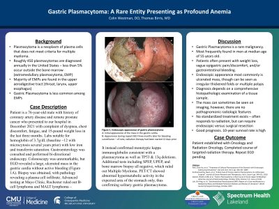
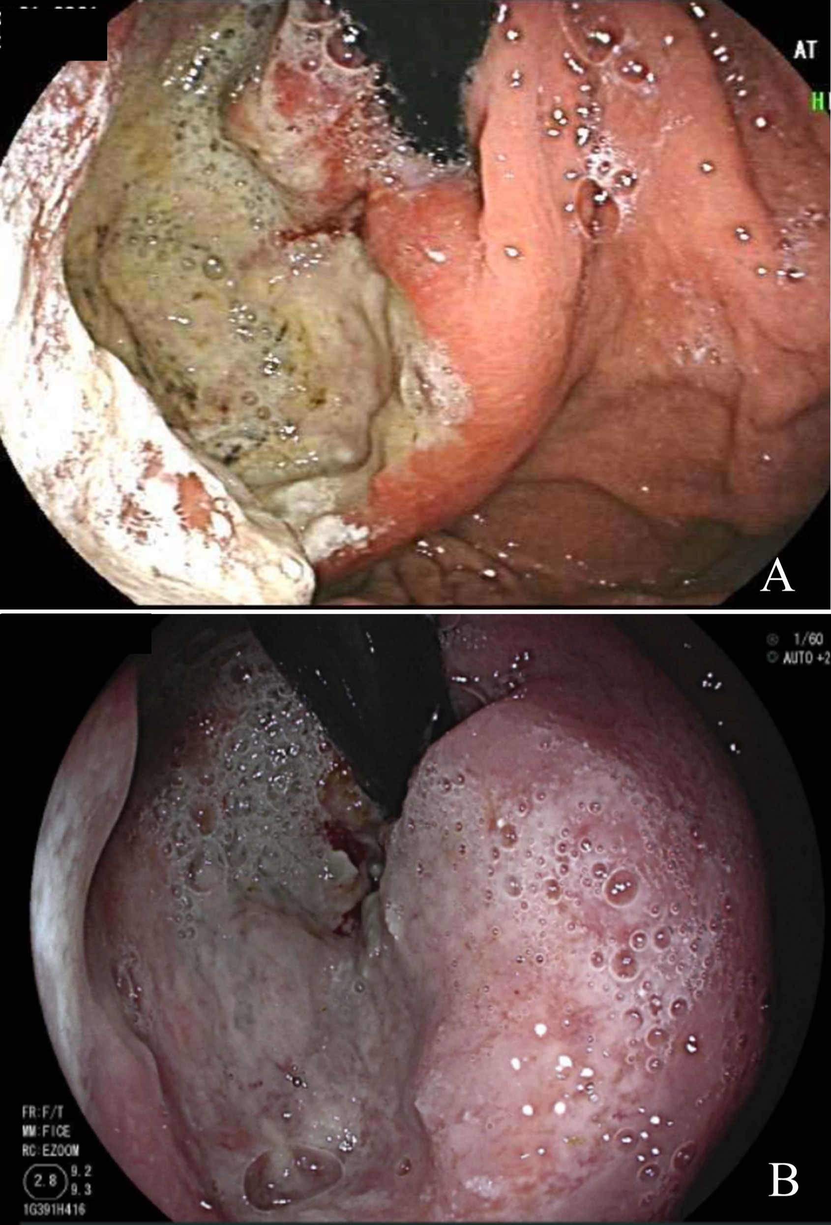
Case Description/Methods: Patient is a 76-year-old male with history of coronary artery disease and remote prostate cancer who presented to our hospital in December 2021 with complaint of shortness of breath, chest discomfort, fatigue, and 15-pound weight loss in the last three months. Labs notable for hemoglobin level of 3.7g/dL (baseline ~12 with microcytosis several years prior) with low iron and transferrin saturation. Gastroenterology was consulted and performed bi-directional endoscopy. Colonoscopy was unremarkable, but EGD showed a large, ulcerated mass in the gastric cardia without active bleeding (Figure 1). Biopsy was obtained, with pathology showing a plasma cell infiltrate. Advanced testing at Mayo Clinic, Rochester ruled out B-cell lymphoma and MALT lymphoma – it instead confirmed monotypic kappa immunoglobulin consistent with a plasmacytoma as well as TP53 & 13q deletions. Additional tests including SPEP, UPEP, and bone marrow biopsy were all negative, which ruled out Multiple Myeloma. PET/CT showed abnormal hypermetabolic activity in the expected area of the stomach only, thus confirming solitary gastric plasmacytoma. Patient completed full course of radiation therapy, with repeat endoscopy pending.
Discussion: Gastric Plasmacytoma is a rare entity. It is most often found in men at median age of 55 years. Patients often present with weight loss, vague epigastric pain/discomfort, and/or gastrointestinal bleeding. Tumor appearance can vary from a simple ulcer to an ulcerated mass. They can occasionally be seen on imaging, but there are no radiographic features to distinguish from other tumors. Diagnosis depends on comprehensive histopathologic examination of a tissue sample. 10-year survival rate is high. There is no standard treatment method. It usually responds well to radiation therapy, but sometimes requires endoscopic or surgical resection. We presented this case due to rarity of the condition and to thus raise awareness for consideration in a differential diagnosis under applicable conditions.

Disclosures:
Colin D. Westman, DO, MS1, Thomas Birris, MD2. C0719 - Gastric Plasmacytoma: A Rare Entity Presenting as Profound Anemia, ACG 2022 Annual Scientific Meeting Abstracts. Charlotte, NC: American College of Gastroenterology.
1Spectrum Health Lakeland, St. Joseph, MI; 2Great Lakes Gastroenterology, St. Joseph, MI
Introduction: Plasmacytoma is defined as a neoplasm of plasma cells in a patient who does not meet criteria for Multiple Myeloma. Roughly 450 plasmacytomas are diagnosed annually in the United States. Extramedullary plasmacytoma, which occurs outside the bone, is rare, accounting for < 5% of all plasma cell neoplasms. They are most commonly found in the upper aerodigestive tract. Gastric Plasmacytoma is an even rarer entity – this case report will detail the discovery and workup of such a lesion.
Case Description/Methods: Patient is a 76-year-old male with history of coronary artery disease and remote prostate cancer who presented to our hospital in December 2021 with complaint of shortness of breath, chest discomfort, fatigue, and 15-pound weight loss in the last three months. Labs notable for hemoglobin level of 3.7g/dL (baseline ~12 with microcytosis several years prior) with low iron and transferrin saturation. Gastroenterology was consulted and performed bi-directional endoscopy. Colonoscopy was unremarkable, but EGD showed a large, ulcerated mass in the gastric cardia without active bleeding (Figure 1). Biopsy was obtained, with pathology showing a plasma cell infiltrate. Advanced testing at Mayo Clinic, Rochester ruled out B-cell lymphoma and MALT lymphoma – it instead confirmed monotypic kappa immunoglobulin consistent with a plasmacytoma as well as TP53 & 13q deletions. Additional tests including SPEP, UPEP, and bone marrow biopsy were all negative, which ruled out Multiple Myeloma. PET/CT showed abnormal hypermetabolic activity in the expected area of the stomach only, thus confirming solitary gastric plasmacytoma. Patient completed full course of radiation therapy, with repeat endoscopy pending.
Discussion: Gastric Plasmacytoma is a rare entity. It is most often found in men at median age of 55 years. Patients often present with weight loss, vague epigastric pain/discomfort, and/or gastrointestinal bleeding. Tumor appearance can vary from a simple ulcer to an ulcerated mass. They can occasionally be seen on imaging, but there are no radiographic features to distinguish from other tumors. Diagnosis depends on comprehensive histopathologic examination of a tissue sample. 10-year survival rate is high. There is no standard treatment method. It usually responds well to radiation therapy, but sometimes requires endoscopic or surgical resection. We presented this case due to rarity of the condition and to thus raise awareness for consideration in a differential diagnosis under applicable conditions.

Figure: Figure 1. Plasmacytoma in the gastric cardia visualized as a large, ulcerated mass at time of discovery (A) and three months later for bleeding surveillance (B).
Disclosures:
Colin Westman indicated no relevant financial relationships.
Thomas Birris indicated no relevant financial relationships.
Colin D. Westman, DO, MS1, Thomas Birris, MD2. C0719 - Gastric Plasmacytoma: A Rare Entity Presenting as Profound Anemia, ACG 2022 Annual Scientific Meeting Abstracts. Charlotte, NC: American College of Gastroenterology.

