Back


Poster Session D - Tuesday Morning
Category: Liver
D0558 - HIV-Associated Iron Overload: A Rare Cause of Elevated Transaminases in Patients With HIV
Tuesday, October 25, 2022
10:00 AM – 12:00 PM ET
Location: Crown Ballroom

Has Audio

Rongyi Sun, BS
University Hospital Cleveland Medical Center
Cleveland, OH
Presenting Author(s)
Rongyi Sun, BS1, Sherin Sallam, MD2
1University Hospital Cleveland Medical Center, Cleveland, OH; 2University Hospitals Cleveland Medical Center/ Case Western Reserve University, Cleveland, OH
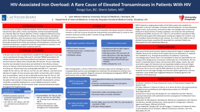
Introduction: Hereditary hemochromatosis can lead to elevated transaminases due to iron deposition in liver. In this case, we discuss a patient with advanced HIV, who in the absence of HFE gene mutation or history of blood transfusions, had drastically elevated ferritin levels and evidence of hemosiderosis in the liver.
Case Description/Methods: A 44-year-old man with a medical history notable for HIV (diagnosed 15 years ago, not on antiretroviral therapy, CD4 count 0, viral load 799000) presented with a transient ischemic attack which resolved after tPA. He was incidentally found to have elevated transaminases and admitted for workup. Patient denied alcohol use for the past 4 years. Physical exam was unremarkable with BMI 17. Labs were notable for ALT 150 u/L, AST 320 u/L, ALP 431 u/L, GGT 1056 u/L, and total serum bilirubin 0.4 mg/dL.
His liver enzymes were within normal limits until 3 months prior to presentation, when he was incidentally found to have ALT 165 u/L, AST 100 u/L, ALP 156 u/L, and total serum bilirubin 0.3 mg/dL. Of note, at that time, the patient had received a course of amoxicillin-clavulanate for pneumonia.
Hepatitis panel was negative for active infection. DILI was suspected due to history of receiving amoxicillin-clavulonate; however, his liver enzymes were still uptrending 3 months after receiving the medication which is uncommon with DILI. AIDS cholangiopathy, a chronic development of biliary strictures caused by infection, was also suspected. MRCP showed no abnormality of biliary system but revealed iron deposition in the liver and spleen consistent with hemosiderosis. Iron studies were then performed showing ferritin 8260 ug/L, serum iron 157 ug/dL, TIBC 272 ug/dL, transferrin saturation 58%. HFE allele genetic testing was negative for mutation, and thus hereditary hemochromatosis was ruled out. Patient was sent home with outpatient hepatology follow-up.
Discussion: HIV commonly targets cells that modulate iron metabolism such as macrophages, and consequently excessive iron accumulates in various organs including liver causing tissue damage, and thus the use of iron chelators has been proposed [1][2]. Our case illustrates the necessity of suspecting HIV-related iron overload in patients with HIV with elevated transaminases of unclear etiology as well as reiterates the potential implications of iron chelation therapy in such cases.
References: [1] Savarino A et al. Cell Biochem Funct. 1999;17(4):279-287. [2] Boelaert JR et al. Infectious Agents and Disease. 1996 Jan;5(1):36-46.
Disclosures:
Rongyi Sun, BS1, Sherin Sallam, MD2. D0558 - HIV-Associated Iron Overload: A Rare Cause of Elevated Transaminases in Patients With HIV, ACG 2022 Annual Scientific Meeting Abstracts. Charlotte, NC: American College of Gastroenterology.
1University Hospital Cleveland Medical Center, Cleveland, OH; 2University Hospitals Cleveland Medical Center/ Case Western Reserve University, Cleveland, OH
Introduction: Hereditary hemochromatosis can lead to elevated transaminases due to iron deposition in liver. In this case, we discuss a patient with advanced HIV, who in the absence of HFE gene mutation or history of blood transfusions, had drastically elevated ferritin levels and evidence of hemosiderosis in the liver.
Case Description/Methods: A 44-year-old man with a medical history notable for HIV (diagnosed 15 years ago, not on antiretroviral therapy, CD4 count 0, viral load 799000) presented with a transient ischemic attack which resolved after tPA. He was incidentally found to have elevated transaminases and admitted for workup. Patient denied alcohol use for the past 4 years. Physical exam was unremarkable with BMI 17. Labs were notable for ALT 150 u/L, AST 320 u/L, ALP 431 u/L, GGT 1056 u/L, and total serum bilirubin 0.4 mg/dL.
His liver enzymes were within normal limits until 3 months prior to presentation, when he was incidentally found to have ALT 165 u/L, AST 100 u/L, ALP 156 u/L, and total serum bilirubin 0.3 mg/dL. Of note, at that time, the patient had received a course of amoxicillin-clavulanate for pneumonia.
Hepatitis panel was negative for active infection. DILI was suspected due to history of receiving amoxicillin-clavulonate; however, his liver enzymes were still uptrending 3 months after receiving the medication which is uncommon with DILI. AIDS cholangiopathy, a chronic development of biliary strictures caused by infection, was also suspected. MRCP showed no abnormality of biliary system but revealed iron deposition in the liver and spleen consistent with hemosiderosis. Iron studies were then performed showing ferritin 8260 ug/L, serum iron 157 ug/dL, TIBC 272 ug/dL, transferrin saturation 58%. HFE allele genetic testing was negative for mutation, and thus hereditary hemochromatosis was ruled out. Patient was sent home with outpatient hepatology follow-up.
Discussion: HIV commonly targets cells that modulate iron metabolism such as macrophages, and consequently excessive iron accumulates in various organs including liver causing tissue damage, and thus the use of iron chelators has been proposed [1][2]. Our case illustrates the necessity of suspecting HIV-related iron overload in patients with HIV with elevated transaminases of unclear etiology as well as reiterates the potential implications of iron chelation therapy in such cases.
References: [1] Savarino A et al. Cell Biochem Funct. 1999;17(4):279-287. [2] Boelaert JR et al. Infectious Agents and Disease. 1996 Jan;5(1):36-46.
Disclosures:
Rongyi Sun indicated no relevant financial relationships.
Sherin Sallam indicated no relevant financial relationships.
Rongyi Sun, BS1, Sherin Sallam, MD2. D0558 - HIV-Associated Iron Overload: A Rare Cause of Elevated Transaminases in Patients With HIV, ACG 2022 Annual Scientific Meeting Abstracts. Charlotte, NC: American College of Gastroenterology.
