Back


Poster Session D - Tuesday Morning
Category: Liver
D0567 - Aden-oh-my!-tosis: A Case of Severe Symptomatic Hepatocellular Adenomatosis
Tuesday, October 25, 2022
10:00 AM – 12:00 PM ET
Location: Crown Ballroom

Has Audio
- JL
John Lee, MD
Walter Reed National Military Medical Center
Bethesda, MD
Presenting Author(s)
Miranda Anderson, DO, Allison Bush, MD, Mark Damiano, MD, John Lee, MD
Walter Reed National Military Medical Center, Bethesda, MD
Introduction: Hepatocellular adenomas (HCA) are benign solid liver lesions often found incidentally on imaging. HCA arise in normal livers, often in women aged 40-50. Large HCA carry risk of malignant transformation and spontaneous rupture.
Hepatocellular adenomatosis is a rare disorder of 10+ HCA. We present a case of severe and symptomatic hepatocellular adenomatosis successfully treated with extensive hepatectomy.
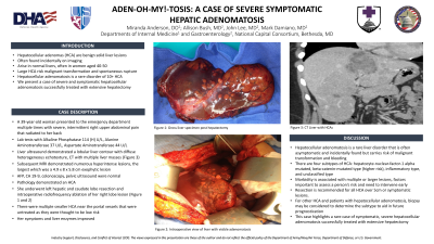
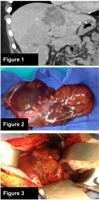
Case Description/Methods: A 39-year-old woman presented to the emergency department multiple times with severe, intermittent right upper abdominal pain that radiated to her back. Lab tests revealed Alkaline Phosphatase 114 (H) U/L, Alanine Aminotransferase 37, Aspartate Aminotransferase 44 U/L. Liver ultrasound demonstrated a lobular liver contour with diffuse heterogeneous echotexture. CT showed multiple liver masses (Figure 1). Subsequent MRI demonstrated numerous hyperintense lesions, the largest was 4.9 x 8 x 5.8 cm exophytic lesion. AFP, CA 19-9, colonoscopy, pelvic ultrasound were normal. Pathology demonstrated a HCA. The patient underwent left hepatic and caudate lobe resection and intraoperative radiofrequency ablation of her right lobe lesion (Figures 2 and 3). There were multiple smaller HCA near the portal vessels that were untreated as they were thought to be low risk. Her symptoms and liver enzymes improved.
Discussion: Hepatocellular adenomatosis is a rare liver disorder that is often asymptomatic and incidentally found but carries risk of malignant transformation and bleeding. There are four subtypes of HCA: hepatocyte-nuclear-factor-1 alpha mutated, beta-catenin-mutated type (higher risk), inflammatory type, and unclassified type. The size and number of lesions is associated with morbidity and warrants consideration for early intervention. Resection is recommended for all HCA over 5cm or symptomatic lesions. For other HCA and patients with hepatocellular adenomatosis, biopsy may be considered to determine the subtype to aid in future prognostication. This case highlights a rare case of symptomatic, severe hepatocellular adenomatosis successfully treated with extensive hepatectomy.

Disclosures:
Miranda Anderson, DO, Allison Bush, MD, Mark Damiano, MD, John Lee, MD. D0567 - Aden-oh-my!-tosis: A Case of Severe Symptomatic Hepatocellular Adenomatosis, ACG 2022 Annual Scientific Meeting Abstracts. Charlotte, NC: American College of Gastroenterology.
Walter Reed National Military Medical Center, Bethesda, MD
Introduction: Hepatocellular adenomas (HCA) are benign solid liver lesions often found incidentally on imaging. HCA arise in normal livers, often in women aged 40-50. Large HCA carry risk of malignant transformation and spontaneous rupture.
Hepatocellular adenomatosis is a rare disorder of 10+ HCA. We present a case of severe and symptomatic hepatocellular adenomatosis successfully treated with extensive hepatectomy.
Case Description/Methods: A 39-year-old woman presented to the emergency department multiple times with severe, intermittent right upper abdominal pain that radiated to her back. Lab tests revealed Alkaline Phosphatase 114 (H) U/L, Alanine Aminotransferase 37, Aspartate Aminotransferase 44 U/L. Liver ultrasound demonstrated a lobular liver contour with diffuse heterogeneous echotexture. CT showed multiple liver masses (Figure 1). Subsequent MRI demonstrated numerous hyperintense lesions, the largest was 4.9 x 8 x 5.8 cm exophytic lesion. AFP, CA 19-9, colonoscopy, pelvic ultrasound were normal. Pathology demonstrated a HCA. The patient underwent left hepatic and caudate lobe resection and intraoperative radiofrequency ablation of her right lobe lesion (Figures 2 and 3). There were multiple smaller HCA near the portal vessels that were untreated as they were thought to be low risk. Her symptoms and liver enzymes improved.
Discussion: Hepatocellular adenomatosis is a rare liver disorder that is often asymptomatic and incidentally found but carries risk of malignant transformation and bleeding. There are four subtypes of HCA: hepatocyte-nuclear-factor-1 alpha mutated, beta-catenin-mutated type (higher risk), inflammatory type, and unclassified type. The size and number of lesions is associated with morbidity and warrants consideration for early intervention. Resection is recommended for all HCA over 5cm or symptomatic lesions. For other HCA and patients with hepatocellular adenomatosis, biopsy may be considered to determine the subtype to aid in future prognostication. This case highlights a rare case of symptomatic, severe hepatocellular adenomatosis successfully treated with extensive hepatectomy.

Figure: Figure 1: CT Liver with HCA. Figure 2: gross liver specimen post hepatectomy. Figure 3: intraoperative view of liver with visible adenomatosis
Disclosures:
Miranda Anderson indicated no relevant financial relationships.
Allison Bush indicated no relevant financial relationships.
Mark Damiano indicated no relevant financial relationships.
John Lee indicated no relevant financial relationships.
Miranda Anderson, DO, Allison Bush, MD, Mark Damiano, MD, John Lee, MD. D0567 - Aden-oh-my!-tosis: A Case of Severe Symptomatic Hepatocellular Adenomatosis, ACG 2022 Annual Scientific Meeting Abstracts. Charlotte, NC: American College of Gastroenterology.
