Back


Poster Session A - Sunday Afternoon
Category: Liver
A0521 - Extensive Hepatic Fibrosis Mimicking Primary Liver Malignancy
Sunday, October 23, 2022
5:00 PM – 7:00 PM ET
Location: Crown Ballroom

Has Audio

Michael Malkowski, MD
St. Elizabeth's Medical Center, Tufts University School of Medicine
Boston, MA
Presenting Author(s)
Michael Malkowski, MD, Ali Niakosari, MD, Tiago Barroso, MD
St. Elizabeth's Medical Center, Tufts University School of Medicine, Boston, MA
Introduction: Liver cirrhosis is a pathophysiological outcome of sustained liver inflammation. Sustained hepatic insults generate a process of fibrogenesis, in which an excessive extracellular matrix deposition replaces normal liver parenchyma. We present a patient with newly diagnosed liver cirrhosis who was found to have a liver mass suggestive of primary liver malignancy on imaging; however, multiple biopsies confirmed the mass to be fibrosis with mild hepatitis.
Case Description/Methods: A 40-year-old male presented for the evaluation of new bilateral lower extremity edema. He reported a long history of alcohol use but otherwise denied prior medical history and no medication use. Laboratory work on admission was pertinent for a total bilirubin of 3.5, AST 162, ALT 57, alkaline phosphatase 176, albumin 3.0, and a non-reactive viral hepatitis panel. CT abdomen/pelvis revealed a heterogeneous liver suggestive of cirrhosis along with a 6.8 cm x 8.7 cm mass with irregular margins situated between the right and left lobes of the liver. On 3-phase MR imaging, the lesion demonstrated progressive enhancement in the portal venous phase and hepatic venous phase, with retention of contrast on 5-minute delayed images [Image 1a, b, c]. Overall, these findings suggested infiltrative cholangiocarcinoma. Additional imaging ruled out multifocal disease, and tumor markers CEA and AFP were unremarkable. Subsequently, a diagnostic laparotomy with liver biopsy was performed. The biopsy demonstrated fibrosis with atypical ductular proliferation, but the presence of low-grade infiltrative cholangiocarcinoma could not be ruled out. To rule out biopsy sampling error, two additional biopsies later that month concurred cirrhotic liver segments with areas of mild hepatitis. Tumor markers CK20 and CDX2 were negative, and no atypical cells were present. It was concluded that this liver lesion represented extensive hepatic fibrosis and mild hepatitis due to ongoing alcohol use, but no malignancy.
Discussion: Liver cirrhosis is a major risk factor for the development of primary liver cancers such as hepatocellular carcinoma and cholangiocarcinoma. Triple-phase CT or MR imaging plays a key role in diagnosing these cancers; however, as demonstrated in our case, certain benign lesions such as liver fibrosis can mimic primary liver malignancy. Improved recognition of these benign lesions is necessary to avoid unnecessary and potentially harmful interventions.

Disclosures:
Michael Malkowski, MD, Ali Niakosari, MD, Tiago Barroso, MD. A0521 - Extensive Hepatic Fibrosis Mimicking Primary Liver Malignancy, ACG 2022 Annual Scientific Meeting Abstracts. Charlotte, NC: American College of Gastroenterology.
St. Elizabeth's Medical Center, Tufts University School of Medicine, Boston, MA
Introduction: Liver cirrhosis is a pathophysiological outcome of sustained liver inflammation. Sustained hepatic insults generate a process of fibrogenesis, in which an excessive extracellular matrix deposition replaces normal liver parenchyma. We present a patient with newly diagnosed liver cirrhosis who was found to have a liver mass suggestive of primary liver malignancy on imaging; however, multiple biopsies confirmed the mass to be fibrosis with mild hepatitis.
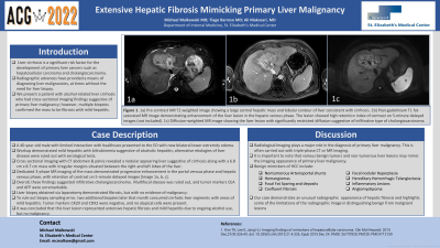
Case Description/Methods: A 40-year-old male presented for the evaluation of new bilateral lower extremity edema. He reported a long history of alcohol use but otherwise denied prior medical history and no medication use. Laboratory work on admission was pertinent for a total bilirubin of 3.5, AST 162, ALT 57, alkaline phosphatase 176, albumin 3.0, and a non-reactive viral hepatitis panel. CT abdomen/pelvis revealed a heterogeneous liver suggestive of cirrhosis along with a 6.8 cm x 8.7 cm mass with irregular margins situated between the right and left lobes of the liver. On 3-phase MR imaging, the lesion demonstrated progressive enhancement in the portal venous phase and hepatic venous phase, with retention of contrast on 5-minute delayed images [Image 1a, b, c]. Overall, these findings suggested infiltrative cholangiocarcinoma. Additional imaging ruled out multifocal disease, and tumor markers CEA and AFP were unremarkable. Subsequently, a diagnostic laparotomy with liver biopsy was performed. The biopsy demonstrated fibrosis with atypical ductular proliferation, but the presence of low-grade infiltrative cholangiocarcinoma could not be ruled out. To rule out biopsy sampling error, two additional biopsies later that month concurred cirrhotic liver segments with areas of mild hepatitis. Tumor markers CK20 and CDX2 were negative, and no atypical cells were present. It was concluded that this liver lesion represented extensive hepatic fibrosis and mild hepatitis due to ongoing alcohol use, but no malignancy.
Discussion: Liver cirrhosis is a major risk factor for the development of primary liver cancers such as hepatocellular carcinoma and cholangiocarcinoma. Triple-phase CT or MR imaging plays a key role in diagnosing these cancers; however, as demonstrated in our case, certain benign lesions such as liver fibrosis can mimic primary liver malignancy. Improved recognition of these benign lesions is necessary to avoid unnecessary and potentially harmful interventions.

Figure: Figure 1: 1a) Pre-contrast MR T2 weighted image showing a large central hepatic mass and lobular contour of liver consistent with cirrhosis. 1b) Post-gadolinium T1 fat-saturated MR image demonstrating enhancement of the liver lesion in the hepatic venous phase. The lesion showed high retention index of contrast on 5-minute delayed images (not included). 1c) Diffusion-weighted MR image showing the liver lesion with significantly restricted diffusion suggestive of infiltrative type of cholangiocarcinoma.
Disclosures:
Michael Malkowski indicated no relevant financial relationships.
Ali Niakosari indicated no relevant financial relationships.
Tiago Barroso indicated no relevant financial relationships.
Michael Malkowski, MD, Ali Niakosari, MD, Tiago Barroso, MD. A0521 - Extensive Hepatic Fibrosis Mimicking Primary Liver Malignancy, ACG 2022 Annual Scientific Meeting Abstracts. Charlotte, NC: American College of Gastroenterology.
