Back


Poster Session D - Tuesday Morning
Category: Colon
D0160 - An Unusual Cause of Rectal Bleeding: Colonic Angiolipoma
Tuesday, October 25, 2022
10:00 AM – 12:00 PM ET
Location: Crown Ballroom

Has Audio
.jpeg.jpg)
Maya Patel, MD, MS
Emory University School of Medicine, University of Arizona College of Medicine-Phoenix
Scottsdale, AZ
Presenting Author(s)
Maya Patel, MD, MS1, Eugene Kim, MD2, Wendi Zhou, MD, PhD2, Shiva Ratuapli, MD2
1Emory University School of Medicine, University of Arizona College of Medicine-Phoenix, Scottsdale, AZ; 2University of Arizona College of Medicine, Phoenix, AZ
Introduction: Angiolipomas are rare, benign, subcutaneous tumors composed of adipose tissue and vascular components. Angiolipomas are extremely rare in the colon, accounting for less than 1% of all colonic benign lesions. While these tumors are typically painful, they may also present with rectal bleeding or positive fecal occult blood test in an otherwise asymptomatic patient. Therefore, it is important to recognize the clinical, radiological, and endoscopic findings associated with these masses.
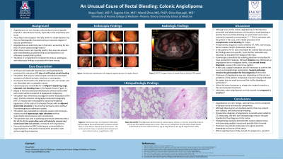
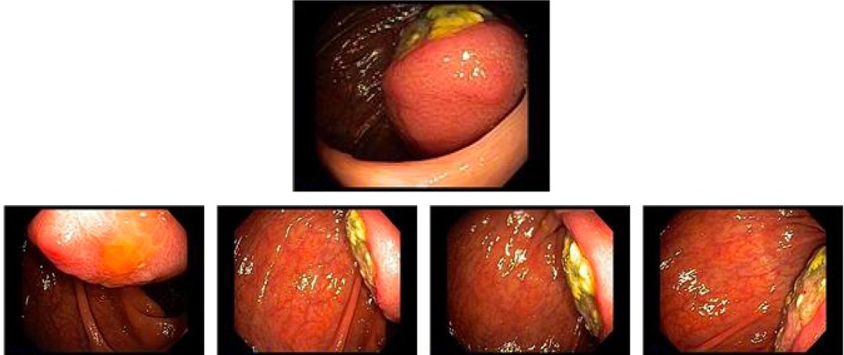
Case Description/Methods: A 49-year-old male with hypertension and hyperlipidemia presented for evaluation of three days of self-limited rectal bleeding. The patient had no prior colonoscopies and denied associated symptoms of abdominal pain, diarrhea, or constipation. On physical examination, the abdomen was soft, non-tender, and non-distended with no palpable masses. Colonoscopy was remarkable for a malignant-appearing, large, ulcerated, non-bleeding mass in the hepatic flexure (Figure 1). Biopsy of the mass demonstrated features of focal active colitis and erosion without evidence of dysplasia or malignancy. The patient was referred to oncology for further evaluation of the mass.
Positron emission tomography-computed tomography (PET-CT) results were remarkable for abnormal thickened appearance of the colon at the hepatic flexure with an adjacent 4.4cm fatty structure in the proximal transverse colon without fluorodeoxyglucose radiotracer uptake. The patient was evaluated by colorectal surgery and received a robotic-assisted laparoscopic right colectomy with an isoperistaltic ileotransverse colon anastomosis. The specimen was sent to pathology and results demonstrated a 4.4 x 4.0 x 4.0 cm protruding mass with glanular mucosa and submucosal fatty cut surfaces, consistent with angiolipoma. Immunohistochemical (IHC) staining was unremarkable for angiomyolipoma. The patient tolerated the procedure well without significant sequelae.
Discussion: Angiolipomas are typically painful yet benign tumors that are rarely found in the gastrointestinal tract. Although abdominal pain, rectal bleeding, and/or positive fecal occult blood testing are commonly reported on presentation, diagnosis is complicated as symptoms tend to be non-specific. Preoperative diagnosis of angiolipoma is possible when aided by CT, endoscopy, and MRI, but surgical pathology remains the gold standard for final diagnosis of the tumor. When angiolipomas are fully excised, the prognosis is excellent.

Disclosures:
Maya Patel, MD, MS1, Eugene Kim, MD2, Wendi Zhou, MD, PhD2, Shiva Ratuapli, MD2. D0160 - An Unusual Cause of Rectal Bleeding: Colonic Angiolipoma, ACG 2022 Annual Scientific Meeting Abstracts. Charlotte, NC: American College of Gastroenterology.
1Emory University School of Medicine, University of Arizona College of Medicine-Phoenix, Scottsdale, AZ; 2University of Arizona College of Medicine, Phoenix, AZ
Introduction: Angiolipomas are rare, benign, subcutaneous tumors composed of adipose tissue and vascular components. Angiolipomas are extremely rare in the colon, accounting for less than 1% of all colonic benign lesions. While these tumors are typically painful, they may also present with rectal bleeding or positive fecal occult blood test in an otherwise asymptomatic patient. Therefore, it is important to recognize the clinical, radiological, and endoscopic findings associated with these masses.
Case Description/Methods: A 49-year-old male with hypertension and hyperlipidemia presented for evaluation of three days of self-limited rectal bleeding. The patient had no prior colonoscopies and denied associated symptoms of abdominal pain, diarrhea, or constipation. On physical examination, the abdomen was soft, non-tender, and non-distended with no palpable masses. Colonoscopy was remarkable for a malignant-appearing, large, ulcerated, non-bleeding mass in the hepatic flexure (Figure 1). Biopsy of the mass demonstrated features of focal active colitis and erosion without evidence of dysplasia or malignancy. The patient was referred to oncology for further evaluation of the mass.
Positron emission tomography-computed tomography (PET-CT) results were remarkable for abnormal thickened appearance of the colon at the hepatic flexure with an adjacent 4.4cm fatty structure in the proximal transverse colon without fluorodeoxyglucose radiotracer uptake. The patient was evaluated by colorectal surgery and received a robotic-assisted laparoscopic right colectomy with an isoperistaltic ileotransverse colon anastomosis. The specimen was sent to pathology and results demonstrated a 4.4 x 4.0 x 4.0 cm protruding mass with glanular mucosa and submucosal fatty cut surfaces, consistent with angiolipoma. Immunohistochemical (IHC) staining was unremarkable for angiomyolipoma. The patient tolerated the procedure well without significant sequelae.
Discussion: Angiolipomas are typically painful yet benign tumors that are rarely found in the gastrointestinal tract. Although abdominal pain, rectal bleeding, and/or positive fecal occult blood testing are commonly reported on presentation, diagnosis is complicated as symptoms tend to be non-specific. Preoperative diagnosis of angiolipoma is possible when aided by CT, endoscopy, and MRI, but surgical pathology remains the gold standard for final diagnosis of the tumor. When angiolipomas are fully excised, the prognosis is excellent.

Figure: Endoscopic identification of malignant-appearing mass in hepatic flexure.
Disclosures:
Maya Patel indicated no relevant financial relationships.
Eugene Kim indicated no relevant financial relationships.
Wendi Zhou indicated no relevant financial relationships.
Shiva Ratuapli indicated no relevant financial relationships.
Maya Patel, MD, MS1, Eugene Kim, MD2, Wendi Zhou, MD, PhD2, Shiva Ratuapli, MD2. D0160 - An Unusual Cause of Rectal Bleeding: Colonic Angiolipoma, ACG 2022 Annual Scientific Meeting Abstracts. Charlotte, NC: American College of Gastroenterology.

