Back


Poster Session A - Sunday Afternoon
Category: Colon
A0148 - A Rare Systemic Illness Presenting With Fever and Abdomen Pain
Sunday, October 23, 2022
5:00 PM – 7:00 PM ET
Location: Crown Ballroom

Has Audio

Anurag Sachan, MBBS, MD
Post Graduate Institute of Medical Education and Research
S.A.S. Nagar, Punjab, India
Presenting Author(s)
Anurag Sachan, MBBS, MD1, Harshal Mandavdhare, MBBS, MD, DM2, Gsrsnk Naidu, MBBS, MD, DM2
1Post Graduate Institute of Medical Education and Research, S.A.S. Nagar, Punjab, India; 2Post Graduate Institute of Medical Education and Research, Chandigarh, Chandigarh, India
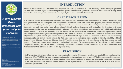
Introduction: Erdheim-Chester disease (ECD) is a rare non-Langerhans cell histiocytic disease. ECD can practically involve any organ system in the body with common organs involved being skeletal system, cardiovascular system and the central nervous system. Rarely, other organs like breast, liver, spleen, kidney, testis and thyroid glands were reported to be involved.
Case Description/Methods: A 51-year-old female presented to our emergency with fever and left upper quadrant pain abdomen of 10 days. Historically, she was symptomatic for last three years with complaints of intermittent fever, knee joints pain, polyuria, nocturia and polydipsia. Contrast enhanced computer tomography (CECT) chest and abdomen showed bilateral pleural and pericardial effusion, thrombosis in splenic vein and right portal vein, splenic infarct, bilateral urothelial thickening with perinephric fat stranding, soft tissue thickening around the aorta and asymmetrical bulky adrenals. PET-CT revealed Flouro-deoxyglucose (FDG) avid ill-defined lesion in the pericardium which was extending into the inter-atrial and atrioventricular septum and FDG avid asymmetrical mural thickening of aorta extending from ascending thoracic aorta up to the infrarenal abdominal aorta. There was presence of bulky tail of pancreas, bulky left adrenal, lytic lesions involving left femoral head, right sacrum and mandible, FDG avid lesions involving meninges, sella and cranial venous sinuses. Tc-99m bone scan showed increased tracer uptake in maxilla, mandible, bilateral forearm bones, bilateral femurs and bilateral tibia. PET guided biopsy from the lytic bone lesion in the right sacrum showed presence of cluster of atypical histiocytes with occasional multinucleated Touton type giant cells. The cells also showed granular cytoplasmic positivity for BRAFV600E, confirming the diagnosis of Erdheim Chester disease (ECD). She was initiated on oral vemurafenib, BRAF inhibitor, at a dose of 240mg twice a day.
Discussion: ECD presenting with splenic infarct has not been described previously. A thorough evaluation and targeted biopsy confirmed the diagnosis. BRAFV600E is the most common mutation noted in these patients, seen in nearly 60% of the patients and the patients with BRAF mutations respond well to vemurafenib, a kinase domain inhibitor of mutant BRAF. Here in, we report a patient of ECD who presented with multisite venous thrombosis and splenic infarct, a rare manifestation of ECD, who was treated successfully with vemurafenib.
Disclosures:
Anurag Sachan, MBBS, MD1, Harshal Mandavdhare, MBBS, MD, DM2, Gsrsnk Naidu, MBBS, MD, DM2. A0148 - A Rare Systemic Illness Presenting With Fever and Abdomen Pain, ACG 2022 Annual Scientific Meeting Abstracts. Charlotte, NC: American College of Gastroenterology.
1Post Graduate Institute of Medical Education and Research, S.A.S. Nagar, Punjab, India; 2Post Graduate Institute of Medical Education and Research, Chandigarh, Chandigarh, India
Introduction: Erdheim-Chester disease (ECD) is a rare non-Langerhans cell histiocytic disease. ECD can practically involve any organ system in the body with common organs involved being skeletal system, cardiovascular system and the central nervous system. Rarely, other organs like breast, liver, spleen, kidney, testis and thyroid glands were reported to be involved.
Case Description/Methods: A 51-year-old female presented to our emergency with fever and left upper quadrant pain abdomen of 10 days. Historically, she was symptomatic for last three years with complaints of intermittent fever, knee joints pain, polyuria, nocturia and polydipsia. Contrast enhanced computer tomography (CECT) chest and abdomen showed bilateral pleural and pericardial effusion, thrombosis in splenic vein and right portal vein, splenic infarct, bilateral urothelial thickening with perinephric fat stranding, soft tissue thickening around the aorta and asymmetrical bulky adrenals. PET-CT revealed Flouro-deoxyglucose (FDG) avid ill-defined lesion in the pericardium which was extending into the inter-atrial and atrioventricular septum and FDG avid asymmetrical mural thickening of aorta extending from ascending thoracic aorta up to the infrarenal abdominal aorta. There was presence of bulky tail of pancreas, bulky left adrenal, lytic lesions involving left femoral head, right sacrum and mandible, FDG avid lesions involving meninges, sella and cranial venous sinuses. Tc-99m bone scan showed increased tracer uptake in maxilla, mandible, bilateral forearm bones, bilateral femurs and bilateral tibia. PET guided biopsy from the lytic bone lesion in the right sacrum showed presence of cluster of atypical histiocytes with occasional multinucleated Touton type giant cells. The cells also showed granular cytoplasmic positivity for BRAFV600E, confirming the diagnosis of Erdheim Chester disease (ECD). She was initiated on oral vemurafenib, BRAF inhibitor, at a dose of 240mg twice a day.
Discussion: ECD presenting with splenic infarct has not been described previously. A thorough evaluation and targeted biopsy confirmed the diagnosis. BRAFV600E is the most common mutation noted in these patients, seen in nearly 60% of the patients and the patients with BRAF mutations respond well to vemurafenib, a kinase domain inhibitor of mutant BRAF. Here in, we report a patient of ECD who presented with multisite venous thrombosis and splenic infarct, a rare manifestation of ECD, who was treated successfully with vemurafenib.
Disclosures:
Anurag Sachan indicated no relevant financial relationships.
Harshal Mandavdhare indicated no relevant financial relationships.
Gsrsnk Naidu indicated no relevant financial relationships.
Anurag Sachan, MBBS, MD1, Harshal Mandavdhare, MBBS, MD, DM2, Gsrsnk Naidu, MBBS, MD, DM2. A0148 - A Rare Systemic Illness Presenting With Fever and Abdomen Pain, ACG 2022 Annual Scientific Meeting Abstracts. Charlotte, NC: American College of Gastroenterology.
