Back


Poster Session D - Tuesday Morning
Category: Small Intestine
D0662 - Malignant Melanoma of Unknown Primary Presenting With Small Intestinal Masses
Tuesday, October 25, 2022
10:00 AM – 12:00 PM ET
Location: Crown Ballroom

Has Audio

Mina Ayad, MD
HCA Florida Aventura Hospital
Aventura, Florida
Presenting Author(s)
Mina Ayad, MD, Ana Martinez, MD, Niel Dave, MD
HCA Florida Aventura Hospital, Aventura, FL
Introduction: GI melanomas are extremely rare and require thorough investigation. Most GI melanomas are metastatic from an oculocutaneous lesion, however, and if not found, are termed melanoma of unknown primary (MUP.) MUP represents only 2% of all melanomas yet it is a major cause of mortality. Common areas of metastasis involve the lymph nodes, small intestines, and subcutaneous sites. We present a case of MUP that developed large abdominal masses within 7 months.
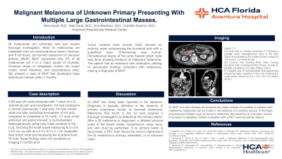
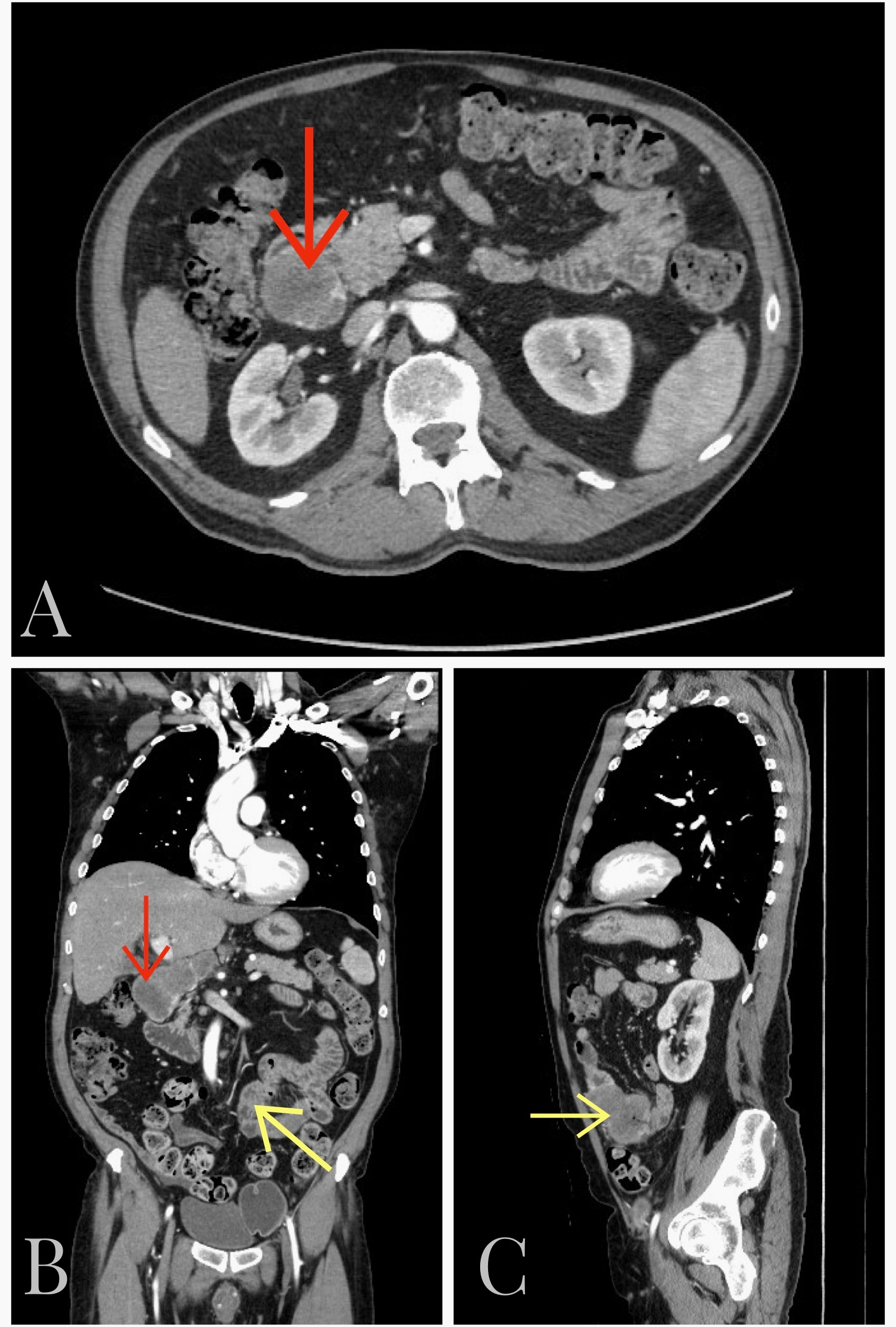
Case Description/Methods: A 68 year old male presented with 1 week of LLQ abdominal pain and constipation. He had undergone a normal colonoscopy 1 year prior. He had normal vitals and labs excluding hemoglobin of 9.5 g/dL, compared to a baseline of 15.2 g/dL. CT scan of the abdomen and pelvis showed: a multi-lobulated heterogeneously enhancing mass centered in the LLQ involving the small bowel measuring 6.8 x 5.6 x 6.5 cm, as well as a 3.5 x 9.0 x 4.1 cm metastatic hilar lymph node encompassing the duodenal bulb. Of note, these findings were not visualized on imaging 7 months prior. Tumor markers were normal. EGD showed an extrinsic mass compressing the duodenal bulb with a superficial ulcer. Colonoscopy was normal. Percutaneous biopsy of the porta hepatis lymph node was done showing evidence of malignant melanoma. The patient had an extensive skin evaluation yielding no skin/ocular findings consistent with melanoma, making a diagnosis of MUP.
Discussion: GI MUP has rarely been reported in the literature. Diagnosis is typically definitive in the absence of primary cutaneous, ocular, or mucosal melanoma. Melanoma first found in the GI tract requires a thorough investigation to determine the primary lesion. After a GI melanoma is diagnosed, a detailed physical exam of the lymph nodes. nasopharynx, eyes, anus, and skin must be performed. If no primary lesion is discovered, a PET scan should be done to determine if the GI melanoma is primary, metastatic, or of unknown origin.

Disclosures:
Mina Ayad, MD, Ana Martinez, MD, Niel Dave, MD. D0662 - Malignant Melanoma of Unknown Primary Presenting With Small Intestinal Masses, ACG 2022 Annual Scientific Meeting Abstracts. Charlotte, NC: American College of Gastroenterology.
HCA Florida Aventura Hospital, Aventura, FL
Introduction: GI melanomas are extremely rare and require thorough investigation. Most GI melanomas are metastatic from an oculocutaneous lesion, however, and if not found, are termed melanoma of unknown primary (MUP.) MUP represents only 2% of all melanomas yet it is a major cause of mortality. Common areas of metastasis involve the lymph nodes, small intestines, and subcutaneous sites. We present a case of MUP that developed large abdominal masses within 7 months.
Case Description/Methods: A 68 year old male presented with 1 week of LLQ abdominal pain and constipation. He had undergone a normal colonoscopy 1 year prior. He had normal vitals and labs excluding hemoglobin of 9.5 g/dL, compared to a baseline of 15.2 g/dL. CT scan of the abdomen and pelvis showed: a multi-lobulated heterogeneously enhancing mass centered in the LLQ involving the small bowel measuring 6.8 x 5.6 x 6.5 cm, as well as a 3.5 x 9.0 x 4.1 cm metastatic hilar lymph node encompassing the duodenal bulb. Of note, these findings were not visualized on imaging 7 months prior. Tumor markers were normal. EGD showed an extrinsic mass compressing the duodenal bulb with a superficial ulcer. Colonoscopy was normal. Percutaneous biopsy of the porta hepatis lymph node was done showing evidence of malignant melanoma. The patient had an extensive skin evaluation yielding no skin/ocular findings consistent with melanoma, making a diagnosis of MUP.
Discussion: GI MUP has rarely been reported in the literature. Diagnosis is typically definitive in the absence of primary cutaneous, ocular, or mucosal melanoma. Melanoma first found in the GI tract requires a thorough investigation to determine the primary lesion. After a GI melanoma is diagnosed, a detailed physical exam of the lymph nodes. nasopharynx, eyes, anus, and skin must be performed. If no primary lesion is discovered, a PET scan should be done to determine if the GI melanoma is primary, metastatic, or of unknown origin.

Figure: Figure 1.0: (A) Axial view of contrast-enhanced CT showing a partially cystic heterogeneous mass in the hilar region encompassing the duodenal bulb measuring 3.5x 9.0x 4.1 cm (Red Arrow). (B) Coronal view showing RUQ mass causing duodenal bulb compression(Red Arrow), as well as partially visualizing LLQ mass (Yellow Arrow.) (C) Sagittal view of multi-lobulated heterogeneously enhancing mass centered in the LLQ involving the small bowel measuring 6.8 x 5.6 x 6.5 cm (Yellow Arrow.)
Disclosures:
Mina Ayad indicated no relevant financial relationships.
Ana Martinez indicated no relevant financial relationships.
Niel Dave indicated no relevant financial relationships.
Mina Ayad, MD, Ana Martinez, MD, Niel Dave, MD. D0662 - Malignant Melanoma of Unknown Primary Presenting With Small Intestinal Masses, ACG 2022 Annual Scientific Meeting Abstracts. Charlotte, NC: American College of Gastroenterology.
