Back


Poster Session D - Tuesday Morning
Category: Liver
D0524 - Hepatic Portal Venous Gas Secondary to Ischemic Gastritis
Tuesday, October 25, 2022
10:00 AM – 12:00 PM ET
Location: Crown Ballroom

Has Audio

Nihal Ijaz Khan, MBBS
Allama Iqbal Medical College
Sarnia, Ontario, Canada
Presenting Author(s)
Nihal Ijaz Khan, MBBS1, Ali Waqar Chaudhry, MD2, Sadaf Raoof, MD3, Syed Hamaad Rahman, DO4, Abu Hurairah, MD3
1Allama Iqbal Medical College, Sarnia, ON, Canada; 2FMH College of Medicine & Dentistry, Lahore, Punjab, Pakistan; 3AdventHealth Orlando, Orlando, FL; 4Methodist Dallas Medical Center, Dallas, TX
Introduction: Hepatic portal venous gas is an uncommon but potentially fatal radiographic finding caused by various disease processes with an overall in-hospital mortality rate of 27.3%. Herein we report a case with an uncommon cause of portal venous gas secondary to severe acute ischemic gastritis associated with gastric pneumatosis.
Case Description/Methods: An 86-year-old male with a past medical history of Parkinsons disease presented with persistent coffee ground emesis for one day. Initial laboratory workup revealed a hemoglobin of 8.9. The patient continued to experience multiple bouts of intractable vomiting unresponsive to antiemetics. He was subsequently intubated and placed on mechanical ventilation for airway protection and transferred to ICU.
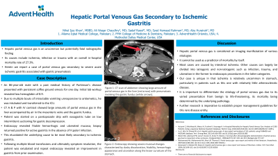
A CT abdomen and pelvis with IV contrast showed large amounts of portal venous gas in the liver accompanied by air in the mesenteric veins adjacent to the stomach and pneumatosis involving the gastric fundus. Patient was started on a Pantoprazole drip and a nasogastric tube was placed with low intermittent suctioning for gastric decompression. Endoscopy revealed friable hemorrhagic and ulcerated mucosa; biopsy returned positive for active gastritis in the absence of H.pylori. This elucidated the underlying cause to be most likely secondary to ischemic gastritis. Following multiple blood transfusions and ultimately symptom resolution, the patient was extubated and repeat endoscopy revealed an improvement in gastritis from prior examination.
Discussion: Hepatic portal venous gas is considered an imaging manifestation of various etiologies and cannot be used as a predictor of mortality by itself. Most cases are caused by intestinal ischemia. Other causes can largely be divided into iatrogenic and non-iatrogenic such as infection, trauma, and ulceration in the former to endoscopic procedures in the latter. Our case is unique in that ischemia is relatively uncommon in stomach, particularly in patients such as this one with relatively little atherosclerotic disease. It is important to differentiate the etiology of portal venous gas due to its varied presentation from benign to life-threatening, its mortality being determined by the underlying pathology.

Disclosures:
Nihal Ijaz Khan, MBBS1, Ali Waqar Chaudhry, MD2, Sadaf Raoof, MD3, Syed Hamaad Rahman, DO4, Abu Hurairah, MD3. D0524 - Hepatic Portal Venous Gas Secondary to Ischemic Gastritis, ACG 2022 Annual Scientific Meeting Abstracts. Charlotte, NC: American College of Gastroenterology.
1Allama Iqbal Medical College, Sarnia, ON, Canada; 2FMH College of Medicine & Dentistry, Lahore, Punjab, Pakistan; 3AdventHealth Orlando, Orlando, FL; 4Methodist Dallas Medical Center, Dallas, TX
Introduction: Hepatic portal venous gas is an uncommon but potentially fatal radiographic finding caused by various disease processes with an overall in-hospital mortality rate of 27.3%. Herein we report a case with an uncommon cause of portal venous gas secondary to severe acute ischemic gastritis associated with gastric pneumatosis.
Case Description/Methods: An 86-year-old male with a past medical history of Parkinsons disease presented with persistent coffee ground emesis for one day. Initial laboratory workup revealed a hemoglobin of 8.9. The patient continued to experience multiple bouts of intractable vomiting unresponsive to antiemetics. He was subsequently intubated and placed on mechanical ventilation for airway protection and transferred to ICU.
A CT abdomen and pelvis with IV contrast showed large amounts of portal venous gas in the liver accompanied by air in the mesenteric veins adjacent to the stomach and pneumatosis involving the gastric fundus. Patient was started on a Pantoprazole drip and a nasogastric tube was placed with low intermittent suctioning for gastric decompression. Endoscopy revealed friable hemorrhagic and ulcerated mucosa; biopsy returned positive for active gastritis in the absence of H.pylori. This elucidated the underlying cause to be most likely secondary to ischemic gastritis. Following multiple blood transfusions and ultimately symptom resolution, the patient was extubated and repeat endoscopy revealed an improvement in gastritis from prior examination.
Discussion: Hepatic portal venous gas is considered an imaging manifestation of various etiologies and cannot be used as a predictor of mortality by itself. Most cases are caused by intestinal ischemia. Other causes can largely be divided into iatrogenic and non-iatrogenic such as infection, trauma, and ulceration in the former to endoscopic procedures in the latter. Our case is unique in that ischemia is relatively uncommon in stomach, particularly in patients such as this one with relatively little atherosclerotic disease. It is important to differentiate the etiology of portal venous gas due to its varied presentation from benign to life-threatening, its mortality being determined by the underlying pathology.

Figure: Figure 1: CT scan of abdomen showing large amount of portal venous gas in the liver (red arrow) with pneumatosis involving the gastric fundus (white arrows).
Figure 2: Endoscopy showing severe mucosal changes characterized by dusky discoloration, friability, hemorrhagic appearance and ulceration along the lesser curvature of the stomach.
Figure 2: Endoscopy showing severe mucosal changes characterized by dusky discoloration, friability, hemorrhagic appearance and ulceration along the lesser curvature of the stomach.
Disclosures:
Nihal Ijaz Khan indicated no relevant financial relationships.
Ali Waqar Chaudhry indicated no relevant financial relationships.
Sadaf Raoof indicated no relevant financial relationships.
Syed Hamaad Rahman indicated no relevant financial relationships.
Abu Hurairah indicated no relevant financial relationships.
Nihal Ijaz Khan, MBBS1, Ali Waqar Chaudhry, MD2, Sadaf Raoof, MD3, Syed Hamaad Rahman, DO4, Abu Hurairah, MD3. D0524 - Hepatic Portal Venous Gas Secondary to Ischemic Gastritis, ACG 2022 Annual Scientific Meeting Abstracts. Charlotte, NC: American College of Gastroenterology.
