Back


Poster Session D - Tuesday Morning
Category: Stomach
D0730 - Gastrointestinal Manifestations of Polyarteritis Nodosa
Tuesday, October 25, 2022
10:00 AM – 12:00 PM ET
Location: Crown Ballroom

Has Audio
.jpg)
Prutha Shah, DO
Albert Einstein Medical Center
Philadelphia, PA
Presenting Author(s)
Prutha Shah, DO, Simone A. Jarrett, MD, Matthew Chan, DO, James Walter, MD
Albert Einstein Medical Center, Philadelphia, PA
Introduction: Polyarteritis Nodosa (PAN) most typically affects medium and small sized vessels. It usually presents with non-specific symptoms of fatigue, myalgia and arthralgias; however, vasculitis is a late presentation. As it commonly involves vital organs such as GI tract and kidneys, delay in treatment can have a high mortality rate due to potential ischemia or micro aneurysmal rupture.
Case Description/Methods: A 50 year old female with history of smoking presented with sharp abdominal pain radiating to left upper quadrant and back. She denied any associated nausea/vomiting or constipation but reported joint pain in toes and fingers, and nodules in her pubic area. Physical exam was remarkable for palpable non-tender raised skin nodule on suprapubic region. Laboratory analysis was significant for elevated rheumatoid factor at 78, mildly elevated ESR 36 and CRP 13.8, complement C3 C4 within normal limits, negative Hepatitis B, C, and negative ANCA and ANA. Biopsy of the suprapubic nodule showed a hemorrhagic nodule with organizing abscess. A CT of abdomen and pelvis revealed a 7.2 x 2.9 x 6.6 cm splenic infarct, perivascular infiltration in the celiac artery extending to common hepatic artery and short gastric artery. The distribution of vessel involvement along the branches of celiac artery suggested PAN. The patient received pulse dose steroids with IV methylprednisone for 3 days. She was discharged on prednisone taper and started on Cyclophosphamide therapy outpatient which resulted in symptomatic improvement.
Discussion: PAN has a predilection for skin, peripheral nerves, GI tract and the kidneys. A classic GI manifestation is mesenteric vasculitis leading to transmural necrotizing inflammation and bowel ischemia. The pathological changes of PAN are limited to the arterial system favoring branch point and thus can be detected with CT or angiography, which is a gold standard for assessment with a sensitivity of 89% and a specificity of 90%. Our case demonstrates a rare complication of PAN vasculitis. Vascular imaging demonstrated celiac trunk involvement that extended into splenic artery leading to subcapsular splenic infarction whereas mesenteric, common hepatic and bilateral renal arteries appeared patent. Timely diagnosis of this condition is important to prevent progression which can include splenic rupture. Our patient experienced symptomatic improvement after glucocorticoids and cyclophosphamide initiation. Follow up vascular imaging is also recommended to monitor for treatment response.

Disclosures:
Prutha Shah, DO, Simone A. Jarrett, MD, Matthew Chan, DO, James Walter, MD. D0730 - Gastrointestinal Manifestations of Polyarteritis Nodosa, ACG 2022 Annual Scientific Meeting Abstracts. Charlotte, NC: American College of Gastroenterology.
Albert Einstein Medical Center, Philadelphia, PA
Introduction: Polyarteritis Nodosa (PAN) most typically affects medium and small sized vessels. It usually presents with non-specific symptoms of fatigue, myalgia and arthralgias; however, vasculitis is a late presentation. As it commonly involves vital organs such as GI tract and kidneys, delay in treatment can have a high mortality rate due to potential ischemia or micro aneurysmal rupture.
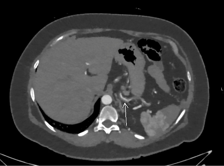
Case Description/Methods: A 50 year old female with history of smoking presented with sharp abdominal pain radiating to left upper quadrant and back. She denied any associated nausea/vomiting or constipation but reported joint pain in toes and fingers, and nodules in her pubic area. Physical exam was remarkable for palpable non-tender raised skin nodule on suprapubic region. Laboratory analysis was significant for elevated rheumatoid factor at 78, mildly elevated ESR 36 and CRP 13.8, complement C3 C4 within normal limits, negative Hepatitis B, C, and negative ANCA and ANA. Biopsy of the suprapubic nodule showed a hemorrhagic nodule with organizing abscess. A CT of abdomen and pelvis revealed a 7.2 x 2.9 x 6.6 cm splenic infarct, perivascular infiltration in the celiac artery extending to common hepatic artery and short gastric artery. The distribution of vessel involvement along the branches of celiac artery suggested PAN. The patient received pulse dose steroids with IV methylprednisone for 3 days. She was discharged on prednisone taper and started on Cyclophosphamide therapy outpatient which resulted in symptomatic improvement.
Discussion: PAN has a predilection for skin, peripheral nerves, GI tract and the kidneys. A classic GI manifestation is mesenteric vasculitis leading to transmural necrotizing inflammation and bowel ischemia. The pathological changes of PAN are limited to the arterial system favoring branch point and thus can be detected with CT or angiography, which is a gold standard for assessment with a sensitivity of 89% and a specificity of 90%. Our case demonstrates a rare complication of PAN vasculitis. Vascular imaging demonstrated celiac trunk involvement that extended into splenic artery leading to subcapsular splenic infarction whereas mesenteric, common hepatic and bilateral renal arteries appeared patent. Timely diagnosis of this condition is important to prevent progression which can include splenic rupture. Our patient experienced symptomatic improvement after glucocorticoids and cyclophosphamide initiation. Follow up vascular imaging is also recommended to monitor for treatment response.

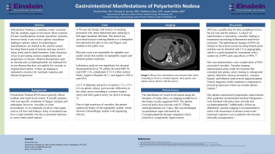
Figure: Ring of low attenuation seen around Celiac artery extending to involve common hepatic, short gastric and splenic artery (as pointed with white arrow)
Disclosures:
Prutha Shah indicated no relevant financial relationships.
Simone Jarrett indicated no relevant financial relationships.
Matthew Chan indicated no relevant financial relationships.
James Walter indicated no relevant financial relationships.
Prutha Shah, DO, Simone A. Jarrett, MD, Matthew Chan, DO, James Walter, MD. D0730 - Gastrointestinal Manifestations of Polyarteritis Nodosa, ACG 2022 Annual Scientific Meeting Abstracts. Charlotte, NC: American College of Gastroenterology.
