Back


Poster Session A - Sunday Afternoon
Category: Colon
A0132 - Two Cases of Lipoma Associated Colo-Colonic Intussusception
Sunday, October 23, 2022
5:00 PM – 7:00 PM ET
Location: Crown Ballroom

Has Audio

Michael Malkowski, MD
St. Elizabeth's Medical Center, Tufts University School of Medicine
Boston, MA
Presenting Author(s)
Michael Malkowski, MD1, Marcel R. Robles, MD2, Sandeep Krishnan, MBBS, PhD3
1St. Elizabeth's Medical Center, Tufts University School of Medicine, Boston, MA; 2Tufts University School of Medicine, St. Elizabeth's Medical Center, Boston, MA; 3St. Elizabeth's Medical Center, Tufts University School of Medicine, Brighton, MA
Introduction: Intussusception in adults is a rare condition. In the pediatric population, 90% of cases are idiopathic, while lead point represents 70-90% of cases in adults. Colo-colonic intussusception (CCI) is most often caused by malignant lead point. We present two cases of CCI caused by large submucosal lipomas.
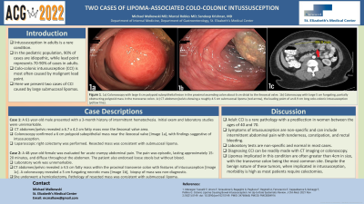
Case Description/Methods: Case 1: A 61-year-old male presented with a 3-month history of intermittent hematochezia. Initial exam and laboratory studies were unremarkable. CT abdomen/pelvis revealed a 6.7 x 4.2 cm fatty mass near the ileocecal valve area. Colonoscopy confirmed a 6 cm polypoid subepithelial mass near the ileocecal valve [Image 1a], with findings suggestive of intussusception. Biopsy of the polyp suggested a hyperplastic polyp. Ultimately, given the persistence of symptoms and mass size, laparoscopic right colectomy was performed. Resected mass was consistent with submucosal lipoma.
Case 2: A 48-year-old female was being evaluated for acute intermittent crampy abdominal pain. The pain was episodic, lasting approximately 15-20 minutes, and diffuse throughout the abdomen. Episodes were self-limited. The patient also endorsed loose stools but otherwise without blood. Laboratory work was unremarkable. CT abdomen/pelvis revealed a 4.5 cm fatty mass within the proximal transverse colon with features suggesting intussusception [Image 1c]. Colonoscopy revealed a 5 cm fungating necrotic mass [Image 1b]; unfortunately, tissue biopsy was nondiagnostic. The patient was initially reluctant to undergo operative management; however, after 5 months of sporadic abdominal cramping, was ultimately amenable for right hemicolectomy, which was curative. Pathology of resected mass was consistent with submucosal lipoma.
Discussion: Adult CCI is a rare pathology with a predilection in women between the ages of 40 and 70. Symptoms of intussusception are non-specific and can include intermittent abdominal pain with tenderness, constipation, and rectal bleeding. Laboratory tests are non-specific and normal in most cases. Diagnosing CCI can be readily made with CT imaging or colonoscopy. Lipomas implicated in this condition are often greater than 4cm in size, with the transverse colon being the most common site. Despite the benign nature of these tumors, when implicated in intussusception, morbidity is high as most patients require colectomies. Both cases highlight differences in symptomatology with this disease.

Disclosures:
Michael Malkowski, MD1, Marcel R. Robles, MD2, Sandeep Krishnan, MBBS, PhD3. A0132 - Two Cases of Lipoma Associated Colo-Colonic Intussusception, ACG 2022 Annual Scientific Meeting Abstracts. Charlotte, NC: American College of Gastroenterology.
1St. Elizabeth's Medical Center, Tufts University School of Medicine, Boston, MA; 2Tufts University School of Medicine, St. Elizabeth's Medical Center, Boston, MA; 3St. Elizabeth's Medical Center, Tufts University School of Medicine, Brighton, MA
Introduction: Intussusception in adults is a rare condition. In the pediatric population, 90% of cases are idiopathic, while lead point represents 70-90% of cases in adults. Colo-colonic intussusception (CCI) is most often caused by malignant lead point. We present two cases of CCI caused by large submucosal lipomas.
Case Description/Methods: Case 1: A 61-year-old male presented with a 3-month history of intermittent hematochezia. Initial exam and laboratory studies were unremarkable. CT abdomen/pelvis revealed a 6.7 x 4.2 cm fatty mass near the ileocecal valve area. Colonoscopy confirmed a 6 cm polypoid subepithelial mass near the ileocecal valve [Image 1a], with findings suggestive of intussusception. Biopsy of the polyp suggested a hyperplastic polyp. Ultimately, given the persistence of symptoms and mass size, laparoscopic right colectomy was performed. Resected mass was consistent with submucosal lipoma.
Case 2: A 48-year-old female was being evaluated for acute intermittent crampy abdominal pain. The pain was episodic, lasting approximately 15-20 minutes, and diffuse throughout the abdomen. Episodes were self-limited. The patient also endorsed loose stools but otherwise without blood. Laboratory work was unremarkable. CT abdomen/pelvis revealed a 4.5 cm fatty mass within the proximal transverse colon with features suggesting intussusception [Image 1c]. Colonoscopy revealed a 5 cm fungating necrotic mass [Image 1b]; unfortunately, tissue biopsy was nondiagnostic. The patient was initially reluctant to undergo operative management; however, after 5 months of sporadic abdominal cramping, was ultimately amenable for right hemicolectomy, which was curative. Pathology of resected mass was consistent with submucosal lipoma.
Discussion: Adult CCI is a rare pathology with a predilection in women between the ages of 40 and 70. Symptoms of intussusception are non-specific and can include intermittent abdominal pain with tenderness, constipation, and rectal bleeding. Laboratory tests are non-specific and normal in most cases. Diagnosing CCI can be readily made with CT imaging or colonoscopy. Lipomas implicated in this condition are often greater than 4cm in size, with the transverse colon being the most common site. Despite the benign nature of these tumors, when implicated in intussusception, morbidity is high as most patients require colectomies. Both cases highlight differences in symptomatology with this disease.

Figure: Figure 1: 1a) Colonoscopy demonstrated a large 6 cm polypoid subepithelial lesion in the proximal ascending colon about 6 cm distal to the ileocecal valve.1b) Colonoscopy demonstrated a large 5 cm fungating, partially obstructing polypoid mass in the transverse colon. 1c) CT abdomen/pelvis showing a roughly 4.5 cm submucosal lipoma (red arrow), the leading point of an 8.9 cm long colo-colonic intussusception (yellow line).
Disclosures:
Michael Malkowski indicated no relevant financial relationships.
Marcel Robles indicated no relevant financial relationships.
Sandeep Krishnan indicated no relevant financial relationships.
Michael Malkowski, MD1, Marcel R. Robles, MD2, Sandeep Krishnan, MBBS, PhD3. A0132 - Two Cases of Lipoma Associated Colo-Colonic Intussusception, ACG 2022 Annual Scientific Meeting Abstracts. Charlotte, NC: American College of Gastroenterology.
