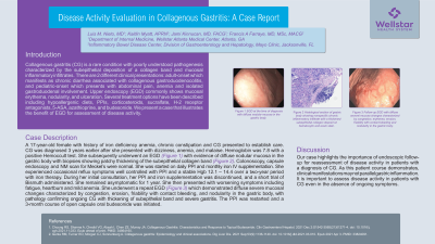
Back


Poster Session D - Tuesday Morning
Category: Stomach
D0734 - Disease Activity Evaluation in Collagenous Gastritis: A Case Report
Tuesday, October 25, 2022
10:00 AM – 12:00 PM ET
Location: Crown Ballroom

Has Audio

Luis M. Nieto, MD
WellStar Atlanta Medical Center
Atlanta, GA
Presenting Author(s)
Luis M. Nieto, MD1, Jami Kinnucan, MD, FACG2, Francis A. Farraye, MD, MSc, MACG3, Kaitlin Myatt, APRN2
1WellStar Atlanta Medical Center, Atlanta, GA; 2Mayo Clinic Florida, Jacksonville, FL; 3Mayo Clinic, Jacksonville, FL
Introduction: Collagenous gastritis (CG) is a rare condition with poorly understood pathogenesis characterized by the subepithelial deposition of a collagen band and mucosal inflammatory infiltrates. There are 2 different clinical presentations: adult-onset which manifests as chronic diarrhea associated with collagenous gastroduodenocolitis, and pediatric-onset which presents with abdominal pain, anemia and isolated gastroduodenal involvement. Upper endoscopy (EGD) commonly shows mucosal erythema, nodularity, and ulceration. Several treatment options have been described including hypoallergenic diets, PPIs, corticosteroids, sucralfate, H-2 receptor antagonists, 5-ASA, azathioprine, and budesonide. We present a case that illustrates the benefit of EGD for assessment of disease activity.
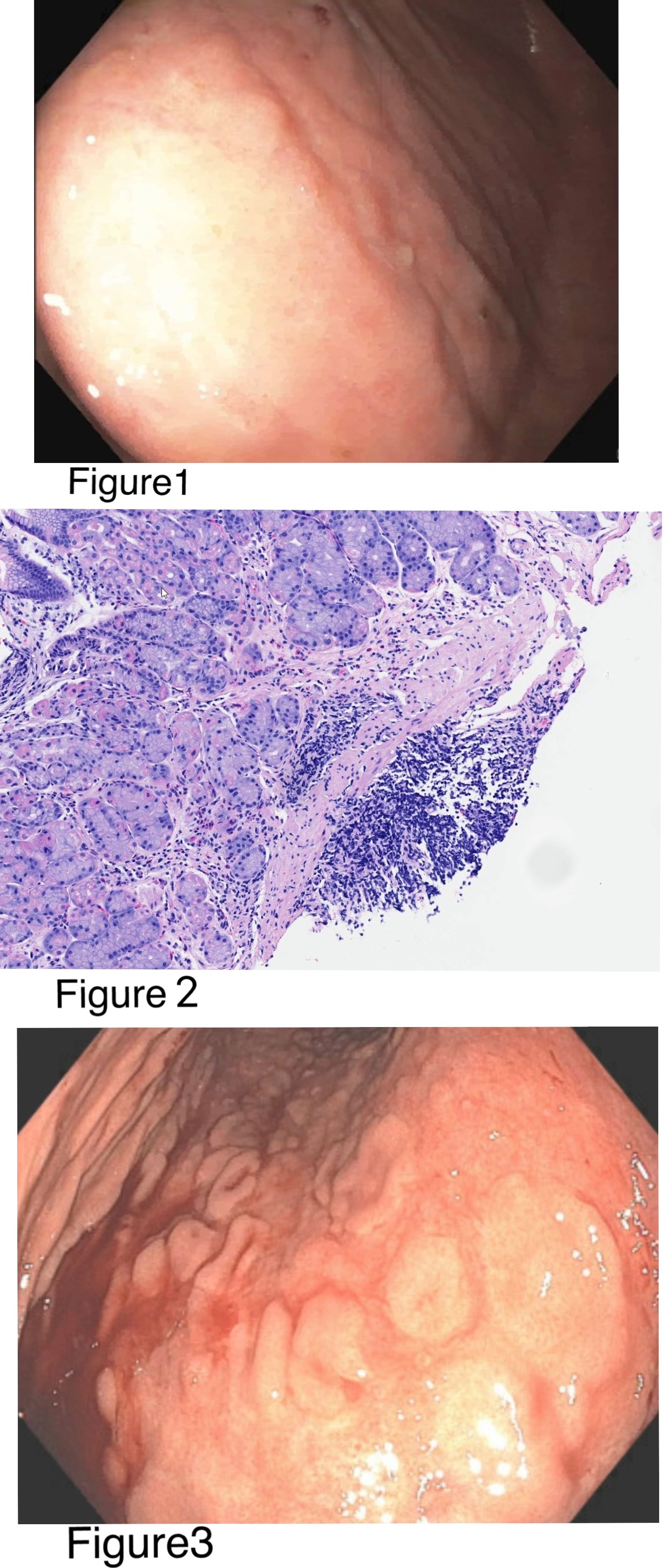
Case Description/Methods: A 17-year-old female with history of iron deficiency anemia, chronic constipation and CG presented to establish care. CG was diagnosed 3 years earlier after she presented with dizziness, anemia, and malaise. Hemoglobin was 7.8 with a positive Hemoccult test. She subsequently underwent an EGD (Figure 1) with evidence of diffuse nodular mucosa in the gastric body with biopsies showing patchy thickening of the subepithelial collagen band (Figure 2). Colonoscopy, capsule endoscopy and NM scan for Meckel’s were normal. She was started on daily PPI and monthly iron IV supplementation. She experienced occasional reflux symptoms well controlled with PPI and a stable Hgb 12.1 – 14.4 over a two-year period with iron therapy. During her initial consultation, Her PPI and iron supplementation was discontinued, and a short trial of Bismuth administered. She remained asymptomatic for 1 year. She then presented with worsening symptoms including fatigue, heartburn and mild anemia. She underwent a repeat EGD (Figure 3) which demonstrated diffuse severe mucosal changes characterized by congestion, erosion, friability with contact bleeding, and nodularity in the gastric body, with pathology confirming ongoing CG with thickening of subepithelial band and severe gastritis. The PPI was restarted and a 3-month course of open capsule oral budesonide was initiated.
Discussion: Our case highlights the importance of endoscopic follow-up for reassessment of disease activity in patients with a diagnosis of CG. As this patient course demonstrates, clinical manifestations may not parallel gastric inflammation. It is important to assess disease activity in patients with CG even in the absence of ongoing symptoms.

Disclosures:
Luis M. Nieto, MD1, Jami Kinnucan, MD, FACG2, Francis A. Farraye, MD, MSc, MACG3, Kaitlin Myatt, APRN2. D0734 - Disease Activity Evaluation in Collagenous Gastritis: A Case Report, ACG 2022 Annual Scientific Meeting Abstracts. Charlotte, NC: American College of Gastroenterology.
1WellStar Atlanta Medical Center, Atlanta, GA; 2Mayo Clinic Florida, Jacksonville, FL; 3Mayo Clinic, Jacksonville, FL
Introduction: Collagenous gastritis (CG) is a rare condition with poorly understood pathogenesis characterized by the subepithelial deposition of a collagen band and mucosal inflammatory infiltrates. There are 2 different clinical presentations: adult-onset which manifests as chronic diarrhea associated with collagenous gastroduodenocolitis, and pediatric-onset which presents with abdominal pain, anemia and isolated gastroduodenal involvement. Upper endoscopy (EGD) commonly shows mucosal erythema, nodularity, and ulceration. Several treatment options have been described including hypoallergenic diets, PPIs, corticosteroids, sucralfate, H-2 receptor antagonists, 5-ASA, azathioprine, and budesonide. We present a case that illustrates the benefit of EGD for assessment of disease activity.
Case Description/Methods: A 17-year-old female with history of iron deficiency anemia, chronic constipation and CG presented to establish care. CG was diagnosed 3 years earlier after she presented with dizziness, anemia, and malaise. Hemoglobin was 7.8 with a positive Hemoccult test. She subsequently underwent an EGD (Figure 1) with evidence of diffuse nodular mucosa in the gastric body with biopsies showing patchy thickening of the subepithelial collagen band (Figure 2). Colonoscopy, capsule endoscopy and NM scan for Meckel’s were normal. She was started on daily PPI and monthly iron IV supplementation. She experienced occasional reflux symptoms well controlled with PPI and a stable Hgb 12.1 – 14.4 over a two-year period with iron therapy. During her initial consultation, Her PPI and iron supplementation was discontinued, and a short trial of Bismuth administered. She remained asymptomatic for 1 year. She then presented with worsening symptoms including fatigue, heartburn and mild anemia. She underwent a repeat EGD (Figure 3) which demonstrated diffuse severe mucosal changes characterized by congestion, erosion, friability with contact bleeding, and nodularity in the gastric body, with pathology confirming ongoing CG with thickening of subepithelial band and severe gastritis. The PPI was restarted and a 3-month course of open capsule oral budesonide was initiated.
Discussion: Our case highlights the importance of endoscopic follow-up for reassessment of disease activity in patients with a diagnosis of CG. As this patient course demonstrates, clinical manifestations may not parallel gastric inflammation. It is important to assess disease activity in patients with CG even in the absence of ongoing symptoms.

Figure: Figure 1 EGD at the time of diagnosis with diffuse nodular mucosa in the gastric body.
Figure 2 Histological section of gastric body showing nonspecific chronic inflammatory infiltrate with a thickened subepithelial collagen deposit on hematoxylin and eosin stain.
Figure 3 Followup EGD with diffuse several mucosa changes characterized by congestion, erythema, erosion, friability with contact bleeding and nodularity in the gastric body.
Disclosures:
Luis Nieto indicated no relevant financial relationships.
Jami Kinnucan: Abbvie – Advisor or Review Panel Member. BMS – Advisor or Review Panel Member. GiH Foundation – Advisor or Review Panel Member. janssen – Advisor or Review Panel Member. Pfizer – Advisor or Review Panel Member, Consultant.
Francis Farraye: Adiso Therapeutics – DSMB. BMS – Advisory Committee/Board Member. Boehringer Ingelheim – Advisory Committee/Board Member. Braintree – Advisory Committee/Board Member. GSK – Advisory Committee/Board Member. IBD Educational Group – Advisory Committee/Board Member. Innovation Pharmaceuticals – Stock Options. Iterative Scopes – Advisory Committee/Board Member. Janssen – Advisory Committee/Board Member. Lilly – DSMB. Pfizer – Advisory Committee/Board Member. Sebela – Advisory Committee/Board Member. Theravance – DSMB.
Kaitlin Myatt: Focus Medical Communications – Presentation Guest Speaker for one presentation.
Luis M. Nieto, MD1, Jami Kinnucan, MD, FACG2, Francis A. Farraye, MD, MSc, MACG3, Kaitlin Myatt, APRN2. D0734 - Disease Activity Evaluation in Collagenous Gastritis: A Case Report, ACG 2022 Annual Scientific Meeting Abstracts. Charlotte, NC: American College of Gastroenterology.
