Back


Poster Session B - Monday Morning
Category: Small Intestine
B0667 - Budesonide in Treating a Patient With Microscopic Enteritis
Monday, October 24, 2022
10:00 AM – 12:00 PM ET
Location: Crown Ballroom

Has Audio
- HA
Hasan Alroobi
Weill Cornell Medicine
New York, NY
Presenting Author(s)
Hasan Alroobi, 1, Malik Mushannen, 1, David Wan, MD2
1Weill Cornell Medicine, New York, NY; 2New York-Presbyterian Hospital/Weill Cornell Medicine, New York, NY
Introduction: Microscopic enteritis is an inflammatory condition in the small bowel that causes chronic diarrhea and bloating. It is associated with gluten intolerance, allergies, drug therapy, inflammatory bowel disease, and autoimmune conditions. Despite the prevalence of risk factors for microscopic enteritis, the understanding of the disease remains limited.
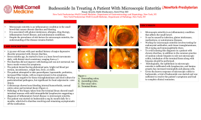
Case Description/Methods: A 33-year-old man with past medical history of major depressive disorder presented with chronic diarrhea. Seven months ago, he started to have 2-5 loose bowel movements daily, with Bristol stool consistency ranging from 4-7. The diarrhea did not improve with fasting and was not nocturnal, but heavy meals worsen his symptoms. The patient denied having abdominal pain, bright red blood per rectum, incontinence, weight loss, fever, or chills. The patient attempted to take pancrelipase, loperamide, probiotics, increased fiber intake, with no improvement in his symptoms. Workup was negative for tissue transglutaminase and stool culture for gastrointestinal pathogens, but significant for fecal calprotectin >200 mcg/g. Colonoscopy showed non-bleeding internal hemorrhoids, normal entire colon and terminal ileum (Figure 1). Pathology of the biopsy taken from the terminal ileum showed small intestinal mucosa with mild intraepithelial lymphocytes suggesting a diagnosis of inflammatory bowel disease vs microscopic enteritis. The patient was started on budesonide 9 mg by mouth daily for 2 months, which led to diarrhea resolving and remaining asymptomatic off the medication.
Discussion: Microscopic enteritis is an inflammatory condition that affects the small bowel. It can be caused by infection, gluten intolerance, medications, or autoimmune diseases. Workup for microscopic enteritis involves testing for endomysial antibodies, anti-tissue transglutaminase, HLA typing and immunoglobulin titers. To avoid missing this diagnosis in a patient with chronic diarrhea, in addition to the common practice of random colonic biopsies to rule out microscopic colitis, intubation of the terminal ileum along with biopsies should be performed. Histologically, the epithelium in microscopic enteritis is infiltrated with lymphocytes, and lamina propria has increased eosinophils and plasma cells. Given the known response of microscopic colitis to budesonide, a trial of budesonide was started and was sufficient to resolve the patient’s symptoms and lead to complete clinical remission.

Disclosures:
Hasan Alroobi, 1, Malik Mushannen, 1, David Wan, MD2. B0667 - Budesonide in Treating a Patient With Microscopic Enteritis, ACG 2022 Annual Scientific Meeting Abstracts. Charlotte, NC: American College of Gastroenterology.
1Weill Cornell Medicine, New York, NY; 2New York-Presbyterian Hospital/Weill Cornell Medicine, New York, NY
Introduction: Microscopic enteritis is an inflammatory condition in the small bowel that causes chronic diarrhea and bloating. It is associated with gluten intolerance, allergies, drug therapy, inflammatory bowel disease, and autoimmune conditions. Despite the prevalence of risk factors for microscopic enteritis, the understanding of the disease remains limited.
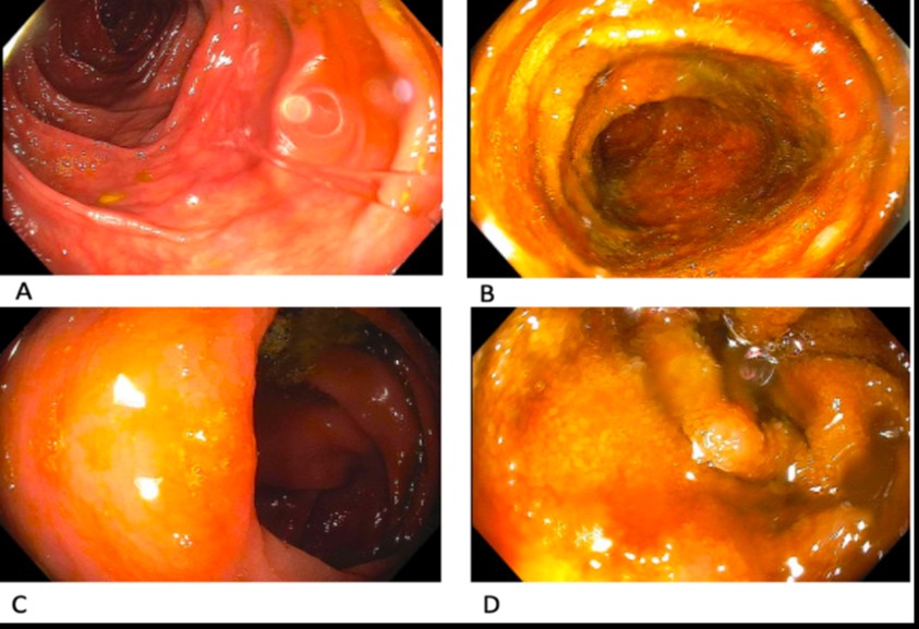
Case Description/Methods: A 33-year-old man with past medical history of major depressive disorder presented with chronic diarrhea. Seven months ago, he started to have 2-5 loose bowel movements daily, with Bristol stool consistency ranging from 4-7. The diarrhea did not improve with fasting and was not nocturnal, but heavy meals worsen his symptoms. The patient denied having abdominal pain, bright red blood per rectum, incontinence, weight loss, fever, or chills. The patient attempted to take pancrelipase, loperamide, probiotics, increased fiber intake, with no improvement in his symptoms. Workup was negative for tissue transglutaminase and stool culture for gastrointestinal pathogens, but significant for fecal calprotectin >200 mcg/g. Colonoscopy showed non-bleeding internal hemorrhoids, normal entire colon and terminal ileum (Figure 1). Pathology of the biopsy taken from the terminal ileum showed small intestinal mucosa with mild intraepithelial lymphocytes suggesting a diagnosis of inflammatory bowel disease vs microscopic enteritis. The patient was started on budesonide 9 mg by mouth daily for 2 months, which led to diarrhea resolving and remaining asymptomatic off the medication.
Discussion: Microscopic enteritis is an inflammatory condition that affects the small bowel. It can be caused by infection, gluten intolerance, medications, or autoimmune diseases. Workup for microscopic enteritis involves testing for endomysial antibodies, anti-tissue transglutaminase, HLA typing and immunoglobulin titers. To avoid missing this diagnosis in a patient with chronic diarrhea, in addition to the common practice of random colonic biopsies to rule out microscopic colitis, intubation of the terminal ileum along with biopsies should be performed. Histologically, the epithelium in microscopic enteritis is infiltrated with lymphocytes, and lamina propria has increased eosinophils and plasma cells. Given the known response of microscopic colitis to budesonide, a trial of budesonide was started and was sufficient to resolve the patient’s symptoms and lead to complete clinical remission.

Figure: Figure 1: A. descending colon; B. ascending colon; C. ileo-cecal valve; D. terminal ileum
Disclosures:
Hasan Alroobi indicated no relevant financial relationships.
Malik Mushannen indicated no relevant financial relationships.
David Wan indicated no relevant financial relationships.
Hasan Alroobi, 1, Malik Mushannen, 1, David Wan, MD2. B0667 - Budesonide in Treating a Patient With Microscopic Enteritis, ACG 2022 Annual Scientific Meeting Abstracts. Charlotte, NC: American College of Gastroenterology.
