Back


Poster Session B - Monday Morning
Category: Stomach
B0706 - Atypical Presentation of Metastatic Breast Cancer
Monday, October 24, 2022
10:00 AM – 12:00 PM ET
Location: Crown Ballroom

Has Audio
.jpg)
Murad H. Ali, MD
University at Buffalo
Buffalo, NY
Presenting Author(s)
Murad H. Ali, MD, Gina M. Sparacino, MD, Mayada Ismail, MD
University at Buffalo, Buffalo, NY
Introduction: Gastrointestinal (GI) sites of metastatic breast cancer (BC) are rare, compared to more frequent sites of bone, lung and brain. Incidence of stomach metastasis from primary tumors is < 1-2%, and according to literature as low as 0.3% from primary BC. The hormone receptor profile of metastatic sites is discordant in up to 15% of cases with triple negative metastatic sites from primary hormone receptor positive tumors. We present a case of metastatic BC to the stomach with tumor discordance.
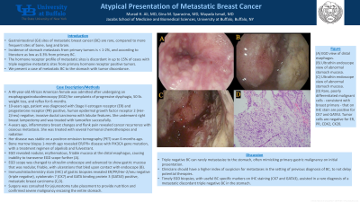
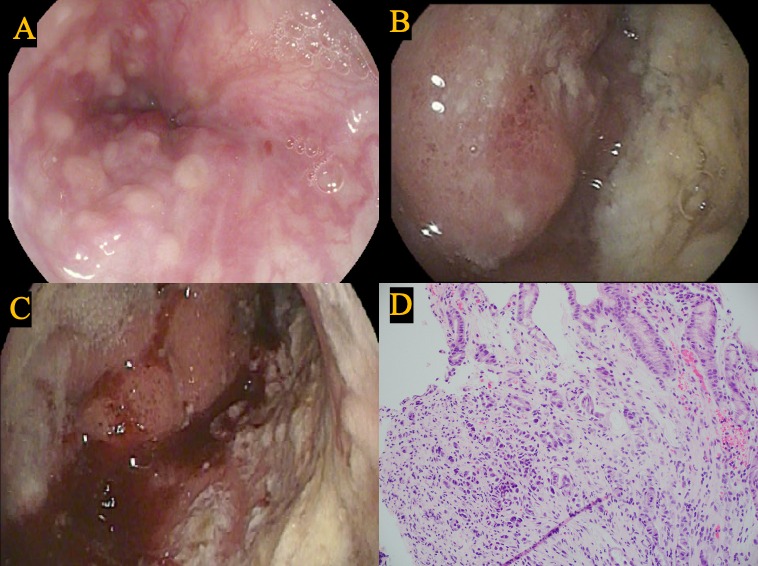
Case Description/Methods: A 49-year-old African American female was admitted after undergoing an esophagogastroduodenoscopy (EGD) for complaints of progressive dysphagia, 50 lb. weight loss, and reflux for 6-months. 10-years ago, patient was diagnosed with Stage II estrogen receptor (ER) and progesterone receptor (PR) positive, human epidermal growth factor receptor 2 (Her-2/neu) negative, invasive ductal carcinoma with lobular features. She underwent right breast lumpectomy and was treated with tamoxifen successfully. 4-years ago, inflammatory breast changes and flank pain revealed cancer recurrence with osseous metastasis. She was treated with several hormonal chemotherapies and radiation, and her disease was stable on a positron emission tomography scan 6-months ago. Bone marrow biopsy 1-month ago revealed ER/PR+ disease with PIK3CA gene mutation, with a treatment regimen of alpelisib and fulvestrant. EGD revealed nodular, erythematous, friable mucosa at the distal esophagus, causing inability to transverse EGD scope further (A). EGD scope was changed to ultraslim endoscope and advanced to show gastric mucosa that was nodular, friable, with ulcerations that bled upon contact with endoscope (B). Immunohistochemistry stain (IHC) of gastric biopsies revealed ER/PR/Her-2/neu negative (triple negative), cytokeratin 7 (CK7) and GATA binding protein 3 (GATA3) positive, metastatic breast carcinoma (C,D). Surgery was consulted for jejunostomy tube placement to provide nutrition and confirmed severe malignancy encasing the entire stomach.
Discussion: Triple negative BC can rarely metastasize to the stomach, often mimicking primary gastric malignancy on initial presentation. Clinicians should have a higher index of suspicion for metastases in the setting of previous diagnosis of BC, to not delay potential therapies. Timely EGD biopsies, with useful BC specific markers on IHC staining (CK7 and GATA3), assisted in a rare diagnosis of a metastatic discordant triple negative BC in the stomach.

Disclosures:
Murad H. Ali, MD, Gina M. Sparacino, MD, Mayada Ismail, MD. B0706 - Atypical Presentation of Metastatic Breast Cancer, ACG 2022 Annual Scientific Meeting Abstracts. Charlotte, NC: American College of Gastroenterology.
University at Buffalo, Buffalo, NY
Introduction: Gastrointestinal (GI) sites of metastatic breast cancer (BC) are rare, compared to more frequent sites of bone, lung and brain. Incidence of stomach metastasis from primary tumors is < 1-2%, and according to literature as low as 0.3% from primary BC. The hormone receptor profile of metastatic sites is discordant in up to 15% of cases with triple negative metastatic sites from primary hormone receptor positive tumors. We present a case of metastatic BC to the stomach with tumor discordance.
Case Description/Methods: A 49-year-old African American female was admitted after undergoing an esophagogastroduodenoscopy (EGD) for complaints of progressive dysphagia, 50 lb. weight loss, and reflux for 6-months. 10-years ago, patient was diagnosed with Stage II estrogen receptor (ER) and progesterone receptor (PR) positive, human epidermal growth factor receptor 2 (Her-2/neu) negative, invasive ductal carcinoma with lobular features. She underwent right breast lumpectomy and was treated with tamoxifen successfully. 4-years ago, inflammatory breast changes and flank pain revealed cancer recurrence with osseous metastasis. She was treated with several hormonal chemotherapies and radiation, and her disease was stable on a positron emission tomography scan 6-months ago. Bone marrow biopsy 1-month ago revealed ER/PR+ disease with PIK3CA gene mutation, with a treatment regimen of alpelisib and fulvestrant. EGD revealed nodular, erythematous, friable mucosa at the distal esophagus, causing inability to transverse EGD scope further (A). EGD scope was changed to ultraslim endoscope and advanced to show gastric mucosa that was nodular, friable, with ulcerations that bled upon contact with endoscope (B). Immunohistochemistry stain (IHC) of gastric biopsies revealed ER/PR/Her-2/neu negative (triple negative), cytokeratin 7 (CK7) and GATA binding protein 3 (GATA3) positive, metastatic breast carcinoma (C,D). Surgery was consulted for jejunostomy tube placement to provide nutrition and confirmed severe malignancy encasing the entire stomach.
Discussion: Triple negative BC can rarely metastasize to the stomach, often mimicking primary gastric malignancy on initial presentation. Clinicians should have a higher index of suspicion for metastases in the setting of previous diagnosis of BC, to not delay potential therapies. Timely EGD biopsies, with useful BC specific markers on IHC staining (CK7 and GATA3), assisted in a rare diagnosis of a metastatic discordant triple negative BC in the stomach.

Figure: (A) EGD view of distal esophagus.
(B) Ultrathin endoscope view of abnormal stomach mucosa.
(C) Ultrathin endoscope view of abnormal stomach mucosa.
(D) Rare, poorly differentiated malignant cells - consistent with breast primary - that on IHC stain are positive for CK7 and GATA3. Tumor cells are negative for ER, PR, CDX2, CK20.
(B) Ultrathin endoscope view of abnormal stomach mucosa.
(C) Ultrathin endoscope view of abnormal stomach mucosa.
(D) Rare, poorly differentiated malignant cells - consistent with breast primary - that on IHC stain are positive for CK7 and GATA3. Tumor cells are negative for ER, PR, CDX2, CK20.
Disclosures:
Murad Ali indicated no relevant financial relationships.
Gina Sparacino indicated no relevant financial relationships.
Mayada Ismail indicated no relevant financial relationships.
Murad H. Ali, MD, Gina M. Sparacino, MD, Mayada Ismail, MD. B0706 - Atypical Presentation of Metastatic Breast Cancer, ACG 2022 Annual Scientific Meeting Abstracts. Charlotte, NC: American College of Gastroenterology.
