Back


Poster Session D - Tuesday Morning
Category: Esophagus
D0254 - Small Cell Neuroendocrine Carcinoma of the Esophagus Presenting as Acute Hematemesis
Tuesday, October 25, 2022
10:00 AM – 12:00 PM ET
Location: Crown Ballroom

Has Audio

Andre Khazak, DO
Mount Sinai Beth Israel
New York, NY
Presenting Author(s)
Andre Khazak, DO1, Carolina Villarroel, MD1, Silpa Yarra, MD1, Minira Aslanova, DO1, Amreen Dinani, MD2
1Mount Sinai Beth Israel, New York, NY; 2Duke University, Durham, NC
Introduction: Esophageal cancer often presents as dysphagia and weight loss and is diagnosed with the help of imaging and endoscopy. Patients with esophageal carcinoma are most commonly found to have either squamous cell carcinoma or esophageal adenocarcinoma. Gastrointestinal small cell neuroendocrine carcinomas account for 0.1 to 1% of all gastrointestinal malignancies and can be seen throughout the GI tract, most commonly noted in the small intestine, appendix, pancreas, and rectum. We present a case of an 85-year-old male with decreasing appetite and 1 day of hematemesis found to have high grade small cell neuroendocrine carcinoma of the esophagus with necrosis.
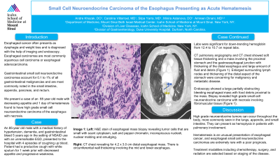
Case Description/Methods: An 85-year old male with a medical history of hypertension, dementia, and gastrointestinal bleed 5 years ago in the setting of NSAID use with an unremarkable EGD, presented to the hospital with 4 episodes of coughing up blood. Patient had a productive cough with white sputum for 1 week with decreased appetite and progressive weakness. On admission, the patient was hemodynamically stable with labs significant for down-trending hemoglobin from 12.4 to 10.7 on repeat labs. CT pulmonary angiography and CT chest for concern for hemoptysis showed soft tissue thickening and a mass involving the proximal stomach and the gastroesophageal junction with thickening of the distal esophagus and large amount of fluid and debris (Figure 1). Furthermore, enlarged surrounding lymph nodes and thickening of the distal aspect of the stomach were concerning for malignancy and metastatic disease. Subsequent endoscopy showed a large partially obstructing bleeding esophageal mass with food debris proximal to the mass. Biopsy revealed high grade small cell neuroendocrine carcinoma with necrosis involving fibromuscular tissue (Figure 1). Given the obstructive nature of presentation and poor prognosis, the family deferred feeding tube placement and elected for inpatient hospice.
Discussion: High grade neuroendocrine tumors can be subdivided into small cell and large cell types and occur throughout the body, more commonly seen in the lungs, appendix, and small intestine. They can present as hemoptysis in patients with pulmonary involvement. Hematemesis is an unusual presentation of esophageal cancer, and esophageal small cell neuroendocrine carcinomas are extremely rare with a poor prognosis given the aggressive nature of the disease. Treatment modalities including chemotherapy, surgery, and radiation are selected based on staging of the disease.

Disclosures:
Andre Khazak, DO1, Carolina Villarroel, MD1, Silpa Yarra, MD1, Minira Aslanova, DO1, Amreen Dinani, MD2. D0254 - Small Cell Neuroendocrine Carcinoma of the Esophagus Presenting as Acute Hematemesis, ACG 2022 Annual Scientific Meeting Abstracts. Charlotte, NC: American College of Gastroenterology.
1Mount Sinai Beth Israel, New York, NY; 2Duke University, Durham, NC
Introduction: Esophageal cancer often presents as dysphagia and weight loss and is diagnosed with the help of imaging and endoscopy. Patients with esophageal carcinoma are most commonly found to have either squamous cell carcinoma or esophageal adenocarcinoma. Gastrointestinal small cell neuroendocrine carcinomas account for 0.1 to 1% of all gastrointestinal malignancies and can be seen throughout the GI tract, most commonly noted in the small intestine, appendix, pancreas, and rectum. We present a case of an 85-year-old male with decreasing appetite and 1 day of hematemesis found to have high grade small cell neuroendocrine carcinoma of the esophagus with necrosis.
Case Description/Methods: An 85-year old male with a medical history of hypertension, dementia, and gastrointestinal bleed 5 years ago in the setting of NSAID use with an unremarkable EGD, presented to the hospital with 4 episodes of coughing up blood. Patient had a productive cough with white sputum for 1 week with decreased appetite and progressive weakness. On admission, the patient was hemodynamically stable with labs significant for down-trending hemoglobin from 12.4 to 10.7 on repeat labs. CT pulmonary angiography and CT chest for concern for hemoptysis showed soft tissue thickening and a mass involving the proximal stomach and the gastroesophageal junction with thickening of the distal esophagus and large amount of fluid and debris (Figure 1). Furthermore, enlarged surrounding lymph nodes and thickening of the distal aspect of the stomach were concerning for malignancy and metastatic disease. Subsequent endoscopy showed a large partially obstructing bleeding esophageal mass with food debris proximal to the mass. Biopsy revealed high grade small cell neuroendocrine carcinoma with necrosis involving fibromuscular tissue (Figure 1). Given the obstructive nature of presentation and poor prognosis, the family deferred feeding tube placement and elected for inpatient hospice.
Discussion: High grade neuroendocrine tumors can be subdivided into small cell and large cell types and occur throughout the body, more commonly seen in the lungs, appendix, and small intestine. They can present as hemoptysis in patients with pulmonary involvement. Hematemesis is an unusual presentation of esophageal cancer, and esophageal small cell neuroendocrine carcinomas are extremely rare with a poor prognosis given the aggressive nature of the disease. Treatment modalities including chemotherapy, surgery, and radiation are selected based on staging of the disease.

Figure: Image 1: Left: H&E stain of esophageal mass biopsy revealing tumor cells that are small with scant cytoplasm, salt and pepper chromatin, inconspicuous nucleoli, nuclear molding and smudging. Right: CT chest revealing for 4.2 x 3.0 cm distal esophageal mass. There is circumferential wall thickening involving the mid and lower esophagus.
Disclosures:
Andre Khazak indicated no relevant financial relationships.
Carolina Villarroel indicated no relevant financial relationships.
Silpa Yarra indicated no relevant financial relationships.
Minira Aslanova indicated no relevant financial relationships.
Amreen Dinani indicated no relevant financial relationships.
Andre Khazak, DO1, Carolina Villarroel, MD1, Silpa Yarra, MD1, Minira Aslanova, DO1, Amreen Dinani, MD2. D0254 - Small Cell Neuroendocrine Carcinoma of the Esophagus Presenting as Acute Hematemesis, ACG 2022 Annual Scientific Meeting Abstracts. Charlotte, NC: American College of Gastroenterology.
