Back


Poster Session B - Monday Morning
Category: Esophagus
B0237 - Incidentally Diagnosed Triple Cancers Within 2 Years: A Novel Case of Rare Metachronous Cancers, Renal Cell Carcinoma, Cholangiocarcinoma, and Squamous Cell Carcinoma of the Gastroesophageal Junction
Monday, October 24, 2022
10:00 AM – 12:00 PM ET
Location: Crown Ballroom

Has Audio
.jpeg.jpg)
Anjeli Manam, BS
University of Illinois College of Medicine
Chicago, IL
Presenting Author(s)
Anjeli Manam, BS1, Krutika Lakhoo, MD2, Itishree Trivedi, MD3
1University of Illinois College of Medicine, Chicago, IL; 2University of Illinois, Chicago, IL; 3University of Illinois Medical Center, Chicago, IL
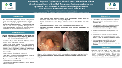
Introduction: The gastrointestinal (GI) tract is involved in many familial cancer syndromes usually involving the colon or stomach. We present a patient with three rare cancers that were detected within a span of 2 years in a single individual: clear cell renal cell carcinoma (ccRCC), cholangiocarcinoma (CCA), and esophageal squamous cell carcinoma (SCC); a combination not previously described in the literature
Case Description/Methods: A 50-year-old male with a history of ccRCC, GERD, and CCA was admitted for evaluation of chronic abdominal pain during chemotherapy for CCA. Regarding his cancer history, ccRCC was incidentally diagnosed 18 months prior on a CT scan for diverticulitis. He underwent a left partial nephrectomy. Surveillance CT imaging 6 months after surgery revealed new incidental liver lesions. Biopsies confirmed metastatic CCA and he started chemotherapy. He noted a 15-year history of episodic abdominal pain and vomiting with no dysphagia or weight loss. Upper endoscopy 16 months prior was normal. He was a former smoker, drank alcohol a few times per month, used marijuana daily, and used cocaine rarely. His family history was significant for multiple cancers at a young age on his maternal side of unknown type. Physical exam showed diffuse abdominal tenderness, worse in the epigastrium without guarding. Labs showed mild anemia, normal BMP/LFTs, and negative H.Pylori stool antigen. Upper endoscopy found nodularity adjacent to the esophagogastric junction (EGJ) with abnormal pit pattern under narrow band imaging (Figure 1). Biopsies confirmed invasive SCC. Staging endoscopic ultrasound showed T3N3 EGJ SCC. He was referred to oncology to discuss treatment options. Genetic testing was positive for BAP1 tumor predisposition syndrome (BAP1-TPDS)
Discussion: A review of published literature did not yield a previous description of this combination of cancers in a single individual. BAP1-TPDS is an uncommon autosomal dominant syndrome caused by germline mutation in a tumor suppressor gene. Patients typically present with melanocytic tumors or malignant mesotheliomas but are at risk for RCC, hepatocellular carcinoma, meningioma and CCA. Literature has not revealed esophageal SCC to be associated. In addition to the incidental nature and timing of diagnosis of all three of these cancers within a short period of 2 years, the EGJ involvement is unusual. This case suggests atypical EGJ-SCC may be considered as part of the BAP1-TPDS tumor spectrum and warrant additional screening for at-risk individuals

Disclosures:
Anjeli Manam, BS1, Krutika Lakhoo, MD2, Itishree Trivedi, MD3. B0237 - Incidentally Diagnosed Triple Cancers Within 2 Years: A Novel Case of Rare Metachronous Cancers, Renal Cell Carcinoma, Cholangiocarcinoma, and Squamous Cell Carcinoma of the Gastroesophageal Junction, ACG 2022 Annual Scientific Meeting Abstracts. Charlotte, NC: American College of Gastroenterology.
1University of Illinois College of Medicine, Chicago, IL; 2University of Illinois, Chicago, IL; 3University of Illinois Medical Center, Chicago, IL
Introduction: The gastrointestinal (GI) tract is involved in many familial cancer syndromes usually involving the colon or stomach. We present a patient with three rare cancers that were detected within a span of 2 years in a single individual: clear cell renal cell carcinoma (ccRCC), cholangiocarcinoma (CCA), and esophageal squamous cell carcinoma (SCC); a combination not previously described in the literature
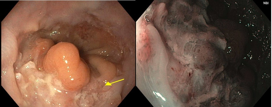
Case Description/Methods: A 50-year-old male with a history of ccRCC, GERD, and CCA was admitted for evaluation of chronic abdominal pain during chemotherapy for CCA. Regarding his cancer history, ccRCC was incidentally diagnosed 18 months prior on a CT scan for diverticulitis. He underwent a left partial nephrectomy. Surveillance CT imaging 6 months after surgery revealed new incidental liver lesions. Biopsies confirmed metastatic CCA and he started chemotherapy. He noted a 15-year history of episodic abdominal pain and vomiting with no dysphagia or weight loss. Upper endoscopy 16 months prior was normal. He was a former smoker, drank alcohol a few times per month, used marijuana daily, and used cocaine rarely. His family history was significant for multiple cancers at a young age on his maternal side of unknown type. Physical exam showed diffuse abdominal tenderness, worse in the epigastrium without guarding. Labs showed mild anemia, normal BMP/LFTs, and negative H.Pylori stool antigen. Upper endoscopy found nodularity adjacent to the esophagogastric junction (EGJ) with abnormal pit pattern under narrow band imaging (Figure 1). Biopsies confirmed invasive SCC. Staging endoscopic ultrasound showed T3N3 EGJ SCC. He was referred to oncology to discuss treatment options. Genetic testing was positive for BAP1 tumor predisposition syndrome (BAP1-TPDS)
Discussion: A review of published literature did not yield a previous description of this combination of cancers in a single individual. BAP1-TPDS is an uncommon autosomal dominant syndrome caused by germline mutation in a tumor suppressor gene. Patients typically present with melanocytic tumors or malignant mesotheliomas but are at risk for RCC, hepatocellular carcinoma, meningioma and CCA. Literature has not revealed esophageal SCC to be associated. In addition to the incidental nature and timing of diagnosis of all three of these cancers within a short period of 2 years, the EGJ involvement is unusual. This case suggests atypical EGJ-SCC may be considered as part of the BAP1-TPDS tumor spectrum and warrant additional screening for at-risk individuals

Figure: Figure 1: Left panel showing abnormal nodular mucosa adjacent to the gastro-esophageal junction under high-definition white light imaging. Right panel showing the same area under narrow band imaging.
Disclosures:
Anjeli Manam indicated no relevant financial relationships.
Krutika Lakhoo indicated no relevant financial relationships.
Itishree Trivedi indicated no relevant financial relationships.
Anjeli Manam, BS1, Krutika Lakhoo, MD2, Itishree Trivedi, MD3. B0237 - Incidentally Diagnosed Triple Cancers Within 2 Years: A Novel Case of Rare Metachronous Cancers, Renal Cell Carcinoma, Cholangiocarcinoma, and Squamous Cell Carcinoma of the Gastroesophageal Junction, ACG 2022 Annual Scientific Meeting Abstracts. Charlotte, NC: American College of Gastroenterology.
