Back


Poster Session E - Tuesday Afternoon
Category: Biliary/Pancreas
E0045 - Hemosuccus Pancreaticus or Just a Bunch of Hocus Pocus
Tuesday, October 25, 2022
3:00 PM – 5:00 PM ET
Location: Crown Ballroom

Has Audio
- DP
Deep Patel, DO
Largo Medical Center
Largo, FL
Presenting Author(s)
Yevgeniya Goltser-Veksler, DO1, Deep Patel, DO2, Nathan Colip, DO2, Meir Mizrahi, MD2, Marc Kudelko, DO2
1Largo Medical Center - HCA Healthcare/USF Morsani College of Medicine, Largo, FL; 2Largo Medical Center, Largo, FL
Introduction: Hemosuccus pancreaticus (HP) is a rare phenomenon defined as bleeding from the pancreatic duct into the gastrointestinal tract via the ampulla of Vater. Many potential causes for HP exist, including pancreatic inflammation, arterial aneurysms, and bariatric surgery. A high degree of clinical suspicion and a multidisciplinary approach is key for early diagnosis and treatment.
Case Description/Methods: 50-year-old female with a history of RYGB, cholecystectomy, pancreatitis, and iron deficiency anemia, was admitted to the intensive care unit after presenting with maroon stools, abdominal pain, and fatigue with a hemoglobin of 4.8. Within the prior 5 months, she had multiple admissions for abdominal pain and GIB and was diagnosed with alcohol-induced pancreatitis, diverticular bleed, anastomotic erosions, peptic ulcer disease, and a Dieulafoy lesion on numerous EGD/colonoscopies. EGD/enteroscopy performed during this admission revealed old blood in the stomach, without active or old blood in the roux or biliopancreatic limbs. Her colonoscopy showed dark tarry stool throughout. Over the subsequent few days, she required a total of 11 units of packed RBCs. Multiple imaging modalities including computed tomography angiography (CTA) and Meckel's diverticulum scan were negative. She experienced worsening abdominal pain, hematochezia and hematemesis, and required emergent intubation. EGD/enteroscopy was performed and revealed fresh blood at the jejuno-jejunal anastomosis, the roux and biliopancreatic limbs, and active bleeding at the ampulla, consistent with HP. Interventional Radiology was contacted immediately, and repeat angiography revealed a 6mm splenic artery pseudoaneurysm. A covered stent was placed, and our patient’s hemoglobin remained stable during the rest of her hospitalization, without further episodes of bleeding or abdominal pain.
Discussion: HP is an obscure, and potentially life-threatening cause of GIB that is important to include in the differential diagnosis. The classic triad includes sporadic bleeding (upper and lower GIB), intermittent abdominal pain, and hyperamylasemia. There appears to be a significant association between HP and pancreatic inflammation, which our patient had. Additionally, our patient’s RYGB anatomy made the ampulla more challenging to reach, delaying the diagnosis. In addition to IR and surgery, advanced endoscopy has presented novel therapeutic options with endoscopic ultrasound, lumen apposing metal stents and fibrin glue/histoacryl adhesives.

Disclosures:
Yevgeniya Goltser-Veksler, DO1, Deep Patel, DO2, Nathan Colip, DO2, Meir Mizrahi, MD2, Marc Kudelko, DO2. E0045 - Hemosuccus Pancreaticus or Just a Bunch of Hocus Pocus, ACG 2022 Annual Scientific Meeting Abstracts. Charlotte, NC: American College of Gastroenterology.
1Largo Medical Center - HCA Healthcare/USF Morsani College of Medicine, Largo, FL; 2Largo Medical Center, Largo, FL
Introduction: Hemosuccus pancreaticus (HP) is a rare phenomenon defined as bleeding from the pancreatic duct into the gastrointestinal tract via the ampulla of Vater. Many potential causes for HP exist, including pancreatic inflammation, arterial aneurysms, and bariatric surgery. A high degree of clinical suspicion and a multidisciplinary approach is key for early diagnosis and treatment.
Case Description/Methods: 50-year-old female with a history of RYGB, cholecystectomy, pancreatitis, and iron deficiency anemia, was admitted to the intensive care unit after presenting with maroon stools, abdominal pain, and fatigue with a hemoglobin of 4.8. Within the prior 5 months, she had multiple admissions for abdominal pain and GIB and was diagnosed with alcohol-induced pancreatitis, diverticular bleed, anastomotic erosions, peptic ulcer disease, and a Dieulafoy lesion on numerous EGD/colonoscopies. EGD/enteroscopy performed during this admission revealed old blood in the stomach, without active or old blood in the roux or biliopancreatic limbs. Her colonoscopy showed dark tarry stool throughout. Over the subsequent few days, she required a total of 11 units of packed RBCs. Multiple imaging modalities including computed tomography angiography (CTA) and Meckel's diverticulum scan were negative. She experienced worsening abdominal pain, hematochezia and hematemesis, and required emergent intubation. EGD/enteroscopy was performed and revealed fresh blood at the jejuno-jejunal anastomosis, the roux and biliopancreatic limbs, and active bleeding at the ampulla, consistent with HP. Interventional Radiology was contacted immediately, and repeat angiography revealed a 6mm splenic artery pseudoaneurysm. A covered stent was placed, and our patient’s hemoglobin remained stable during the rest of her hospitalization, without further episodes of bleeding or abdominal pain.
Discussion: HP is an obscure, and potentially life-threatening cause of GIB that is important to include in the differential diagnosis. The classic triad includes sporadic bleeding (upper and lower GIB), intermittent abdominal pain, and hyperamylasemia. There appears to be a significant association between HP and pancreatic inflammation, which our patient had. Additionally, our patient’s RYGB anatomy made the ampulla more challenging to reach, delaying the diagnosis. In addition to IR and surgery, advanced endoscopy has presented novel therapeutic options with endoscopic ultrasound, lumen apposing metal stents and fibrin glue/histoacryl adhesives.

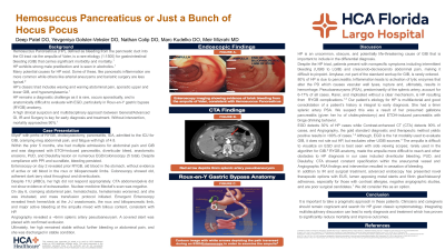
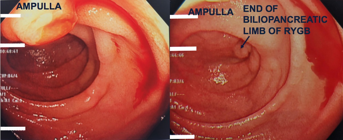
Figure: Enteroscopy imaging showing evidence of brisk bleeding from the ampulla of Vater, consistent with hemosuccus pancreaticus
Disclosures:
Yevgeniya Goltser-Veksler indicated no relevant financial relationships.
Deep Patel indicated no relevant financial relationships.
Nathan Colip indicated no relevant financial relationships.
Meir Mizrahi: Boston Scientific – Consultant. Cook Medical – Consultant. FujiFilm – Consultant. Olympus – Consultant.
Marc Kudelko indicated no relevant financial relationships.
Yevgeniya Goltser-Veksler, DO1, Deep Patel, DO2, Nathan Colip, DO2, Meir Mizrahi, MD2, Marc Kudelko, DO2. E0045 - Hemosuccus Pancreaticus or Just a Bunch of Hocus Pocus, ACG 2022 Annual Scientific Meeting Abstracts. Charlotte, NC: American College of Gastroenterology.
