Back


Poster Session E - Tuesday Afternoon
Category: Biliary/Pancreas
E0061 - Solid Pseudo-Papillary Pancreatic Tumor With a Rare Advanced Metastatic Presentation
Tuesday, October 25, 2022
3:00 PM – 5:00 PM ET
Location: Crown Ballroom

Has Audio

Kosisochukwu J. Ezeh, MD
Jersey City Medical Center
Jersey City, New Jersey
Presenting Author(s)
Kosisochukwu J. Ezeh, MD, Yasir Rajwana, MD, Youssef Botros, MD
Jersey City Medical Center, Jersey City, NJ
Introduction: Solid pseudopapillary tumor (SPT) of the pancreas is a rare neoplasm, usually characterized by a well encapsulated mass, with low malignant potential. It occurs predominantly in young females, in the third decade of life, accounting for about 1% of all tumors of the pancreas mainly located in the body and tail. SPT usually exhibits benign behavior but we present a case of a young female with advanced metastatic disease on presentation.
Case Description/Methods: A 32-year-old female with history of gastritis presents with stabbing, intermittent epigastric pain radiating to the back. In the past, she had presented to the emergency department for gastritis. Vitals notable for low-grade fever and tachycardia. She had no prior colonoscopy/EGD. Family history significant for colon cancer in dad at age of 70. Physical exam was remarkable for epigastric tenderness. Labs showed normal complete blood count, chemistry, liver function tests, lipase. Liver function test were mildly elevated with AST 40 and alkaline phosphatase 131.
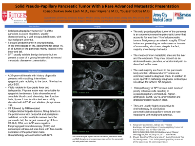
CT followed by MRI revealed multiple bilobar hepatic lesions, filling defects in the portal veins with periportal and perisplenic collateral, complex multiple masses from the pancreatic tail, the largest measuring 14.9cm. CA19-9, CEA, and AFP were negative. Esophagogastroduodenoscopy (EGD) and endoscopic ultrasound was done with fine-needle aspiration of the pancreatic mass. Histopathology was consistent with SPT.
Discussion: The solid pseudopapillary tumor of the pancreas is an uncommon exocrine pancreatic tumor that accounts for less than 1% of all pancreatic tumors. Malignancy can arise in roughly 15% of instances, presenting as metastases or invasion of surrounding structures, despite the fact that majority show benign behavior.
The most common metastatic sites are the liver and the omentum. They may present as an abdominal mass, jaundice, or abdominal pain as described in this case.
The vast majority are found in the pancreatic body and tail. Ultrasound or CT scans are commonly used to diagnose them. In addition to a pre-operative pathology diagnosis, endoscopic US allows for further FNA biopsies. Histopathology of SPT reveals solid nests of poorly cohesive cells resulting in a pseudopapillary architecture. Alpha1-antitrypsin, CD56, CD10, and Vimentin are characteristically found in them. They are usually highly responsive to chemotherapy. In conclusion, pancreatic pseudopapillary tumors are rare neoplasms with malignant potential.

Disclosures:
Kosisochukwu J. Ezeh, MD, Yasir Rajwana, MD, Youssef Botros, MD. E0061 - Solid Pseudo-Papillary Pancreatic Tumor With a Rare Advanced Metastatic Presentation, ACG 2022 Annual Scientific Meeting Abstracts. Charlotte, NC: American College of Gastroenterology.
Jersey City Medical Center, Jersey City, NJ
Introduction: Solid pseudopapillary tumor (SPT) of the pancreas is a rare neoplasm, usually characterized by a well encapsulated mass, with low malignant potential. It occurs predominantly in young females, in the third decade of life, accounting for about 1% of all tumors of the pancreas mainly located in the body and tail. SPT usually exhibits benign behavior but we present a case of a young female with advanced metastatic disease on presentation.
Case Description/Methods: A 32-year-old female with history of gastritis presents with stabbing, intermittent epigastric pain radiating to the back. In the past, she had presented to the emergency department for gastritis. Vitals notable for low-grade fever and tachycardia. She had no prior colonoscopy/EGD. Family history significant for colon cancer in dad at age of 70. Physical exam was remarkable for epigastric tenderness. Labs showed normal complete blood count, chemistry, liver function tests, lipase. Liver function test were mildly elevated with AST 40 and alkaline phosphatase 131.
CT followed by MRI revealed multiple bilobar hepatic lesions, filling defects in the portal veins with periportal and perisplenic collateral, complex multiple masses from the pancreatic tail, the largest measuring 14.9cm. CA19-9, CEA, and AFP were negative. Esophagogastroduodenoscopy (EGD) and endoscopic ultrasound was done with fine-needle aspiration of the pancreatic mass. Histopathology was consistent with SPT.
Discussion: The solid pseudopapillary tumor of the pancreas is an uncommon exocrine pancreatic tumor that accounts for less than 1% of all pancreatic tumors. Malignancy can arise in roughly 15% of instances, presenting as metastases or invasion of surrounding structures, despite the fact that majority show benign behavior.
The most common metastatic sites are the liver and the omentum. They may present as an abdominal mass, jaundice, or abdominal pain as described in this case.
The vast majority are found in the pancreatic body and tail. Ultrasound or CT scans are commonly used to diagnose them. In addition to a pre-operative pathology diagnosis, endoscopic US allows for further FNA biopsies. Histopathology of SPT reveals solid nests of poorly cohesive cells resulting in a pseudopapillary architecture. Alpha1-antitrypsin, CD56, CD10, and Vimentin are characteristically found in them. They are usually highly responsive to chemotherapy. In conclusion, pancreatic pseudopapillary tumors are rare neoplasms with malignant potential.

Figure: MRI showing multiple hepatic lesions as well as pancreatic mass and endoscopic ultrasound illustrating heterogenous mass within the pancreatic tail with portal vein invasion
Disclosures:
Kosisochukwu Ezeh indicated no relevant financial relationships.
Yasir Rajwana indicated no relevant financial relationships.
Youssef Botros indicated no relevant financial relationships.
Kosisochukwu J. Ezeh, MD, Yasir Rajwana, MD, Youssef Botros, MD. E0061 - Solid Pseudo-Papillary Pancreatic Tumor With a Rare Advanced Metastatic Presentation, ACG 2022 Annual Scientific Meeting Abstracts. Charlotte, NC: American College of Gastroenterology.
