Back


Poster Session B - Monday Morning
Category: GI Bleeding
B0344 - Are We Overlooking Small Bowel Diverticular Bleeds? Emergency Partial Jejunectomy After Massive Jejunal Diverticular Bleed
Monday, October 24, 2022
10:00 AM – 12:00 PM ET
Location: Crown Ballroom

Has Audio

Mustafa Abdulsada, MD
Mather Hospital
Port Jefferson, NY
Presenting Author(s)
Mustafa Abdulsada, MD1, Lindsey Jones, MD1, Karina Fatakhova, MD2, Pratik Patel, MD2, Robert Baranowski, MD3
1Mather Hospital, Port Jefferson, NY; 2Mather Hospital, Northwell Health, Port Jefferson, NY; 3Northwell Health, East Setauket, NY
Introduction: The incidence of small bowel diverticula based on autopsy and radiographic results is reported to range from 1-3%. Small bowel diverticula are most commonly located in the jejunum. As with colonic diverticula, it is a disease of the elderly. Severe life-threatening complications of small bowel diverticula including perforation, volvulus, hemorrhage, diverticulitis, and intestinal obstruction have been reported in 10-30% of cases. We present a difficult case of jejunal diverticular bleed requiring emergency surgery.
Case Description/Methods: An 80-year-old male presented to the emergency room three times over the period of 14 days complaining of bright red blood per rectum. He underwent CT scan of his abdomen and pelvis with contrast and colonoscopy during his first two presentations. Imaging was unremarkable with colonoscopy revealing blood throughout the colon, with no evidence of active bleed and a non-bloody terminal ileum. Six days after his second hospital discharge, the patient presented to the ED complaining of dizziness and melena. He was hypotensive and anemic (Hb 6.7 mg/dl). Nuclear tagged RBC scan was significant for focal labeled RBC accumulation in the left upper to the middle quadrant of the abdomen, with activity increasing in intensity over time and dissipating as the study progressed, indicating an active bleeding site, likely in the proximal jejunum. Emergency laparotomy was performed, multiple wide mouth bleeding diverticula within 60 cm of proximal jejunum were found. The affected segment of the jejunum was resected and a primary anastomosis was created. The patient recovered well post op and was eventually discharged to subacute rehabilitation.
Discussion: Although small bowel diverticular bleeds are rarely encountered, it should remain under the differential, especially if a source of bleed is not identified on upper endoscopy (EGD) or colonoscopy. As in our case, repeat procedures may be performed resulting in a delay in diagnosis. Radiologic bleeding scans such as CT angiogram and/or RBC scintigraphy could help diagnose small bowel diverticular bleeding, preventing unnecessary workup. Patients presenting with repeat or persistent brisk rectal bleeding despite negative EGD/Colonoscopy should be considered for imaging to rule out small bowel diverticular bleed.

Disclosures:
Mustafa Abdulsada, MD1, Lindsey Jones, MD1, Karina Fatakhova, MD2, Pratik Patel, MD2, Robert Baranowski, MD3. B0344 - Are We Overlooking Small Bowel Diverticular Bleeds? Emergency Partial Jejunectomy After Massive Jejunal Diverticular Bleed, ACG 2022 Annual Scientific Meeting Abstracts. Charlotte, NC: American College of Gastroenterology.
1Mather Hospital, Port Jefferson, NY; 2Mather Hospital, Northwell Health, Port Jefferson, NY; 3Northwell Health, East Setauket, NY
Introduction: The incidence of small bowel diverticula based on autopsy and radiographic results is reported to range from 1-3%. Small bowel diverticula are most commonly located in the jejunum. As with colonic diverticula, it is a disease of the elderly. Severe life-threatening complications of small bowel diverticula including perforation, volvulus, hemorrhage, diverticulitis, and intestinal obstruction have been reported in 10-30% of cases. We present a difficult case of jejunal diverticular bleed requiring emergency surgery.
Case Description/Methods: An 80-year-old male presented to the emergency room three times over the period of 14 days complaining of bright red blood per rectum. He underwent CT scan of his abdomen and pelvis with contrast and colonoscopy during his first two presentations. Imaging was unremarkable with colonoscopy revealing blood throughout the colon, with no evidence of active bleed and a non-bloody terminal ileum. Six days after his second hospital discharge, the patient presented to the ED complaining of dizziness and melena. He was hypotensive and anemic (Hb 6.7 mg/dl). Nuclear tagged RBC scan was significant for focal labeled RBC accumulation in the left upper to the middle quadrant of the abdomen, with activity increasing in intensity over time and dissipating as the study progressed, indicating an active bleeding site, likely in the proximal jejunum. Emergency laparotomy was performed, multiple wide mouth bleeding diverticula within 60 cm of proximal jejunum were found. The affected segment of the jejunum was resected and a primary anastomosis was created. The patient recovered well post op and was eventually discharged to subacute rehabilitation.
Discussion: Although small bowel diverticular bleeds are rarely encountered, it should remain under the differential, especially if a source of bleed is not identified on upper endoscopy (EGD) or colonoscopy. As in our case, repeat procedures may be performed resulting in a delay in diagnosis. Radiologic bleeding scans such as CT angiogram and/or RBC scintigraphy could help diagnose small bowel diverticular bleeding, preventing unnecessary workup. Patients presenting with repeat or persistent brisk rectal bleeding despite negative EGD/Colonoscopy should be considered for imaging to rule out small bowel diverticular bleed.

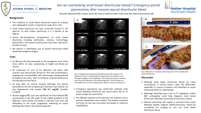
Figure: A) Beginning at approximately 88 minutes after injection of a radiotracer, there is focal labeled red blood cell accumulation in the left upper quadrant of the abdomen. This activity increases in intensity over time and dissipates as the study progresses. These findings are compatible with an active bleeding site, likely in the proximal jejunum. B and C) 60 cm of resected proximal jejunum with multiple wide mouth diverticula.
Disclosures:
Mustafa Abdulsada indicated no relevant financial relationships.
Lindsey Jones indicated no relevant financial relationships.
Karina Fatakhova indicated no relevant financial relationships.
Pratik Patel indicated no relevant financial relationships.
Robert Baranowski indicated no relevant financial relationships.
Mustafa Abdulsada, MD1, Lindsey Jones, MD1, Karina Fatakhova, MD2, Pratik Patel, MD2, Robert Baranowski, MD3. B0344 - Are We Overlooking Small Bowel Diverticular Bleeds? Emergency Partial Jejunectomy After Massive Jejunal Diverticular Bleed, ACG 2022 Annual Scientific Meeting Abstracts. Charlotte, NC: American College of Gastroenterology.
