Back


Poster Session B - Monday Morning
Category: General Endoscopy
B0294 - Think "Inside the Loop:" A Novel Method for Repositioning a Migrated G-J Tube
Monday, October 24, 2022
10:00 AM – 12:00 PM ET
Location: Crown Ballroom

Has Audio
.jpg)
Carol Monis, MD
HCA Healthcare, Riverside Community Hospital
Riverside, CA
Presenting Author(s)
Carol Monis, MD1, Fadi Totah, DO1, Mladen Jecmenica, MD2
1HCA Healthcare, Riverside Community Hospital, Riverside, CA; 2Riverside Community Hospital, Riverside, CA
Introduction: Gastrojejunostomy tube (G-J tube) migration is common in patients requiring long term percutaneous enteral feeding. These patients often present with forceful vomiting and decreased PO intake. This case discusses a method for repositioning a migrated G-J tube using an endoloop (detachable snare device) and Resolution Clips.
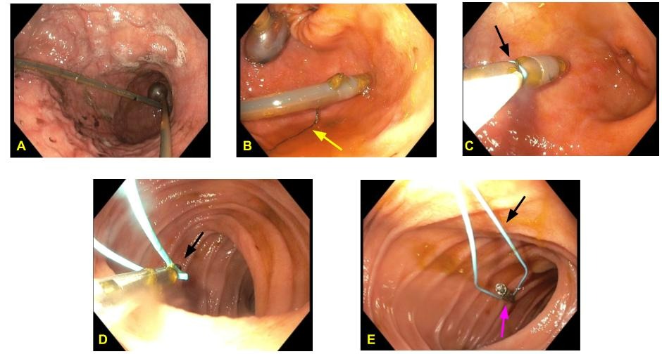
Case Description/Methods: A 26-year-old female with a history of cystic fibrosis (CF), chronic malnutrition requiring enteral feeds, CF-related liver disease and variceal bleeding presented with 5 days of abdominal pain and coffee ground emesis. Initially, the patient was tachycardic and vomiting intermittently. Bright red blood was visible in the emesis bag. Physical exam was notable for epigastric tenderness without peritoneal signs. The skin around the G-J tube insertion site was non-tender with no signs of infection. CT of the abdomen revealed gastrosplenic varices and appropriate positioning of the enteral tube. There was no evidence of GI perforation or obstruction. An EGD demonstrated a patent gastrostomy tube in the gastric body. The jejunal tail was coiled up against the inflamed lumen of the stomach. The suture at the tip of the tail had degraded, dislodging the tube from its original position along the jejunal wall. There were blood clots in the body of the stomach but no evidence of active bleeding. Given the suspicion that the migrated feeding tube was the cause of the patient’s symptoms, a decision was made to reposition it. An endoloop was tied around the tip of the G-J tube to serve as a substitute for the suture. A cold snare was then used to secure the endoloop and drag the tail into the jejunum. The endoloop was secured to the intestinal wall using two Resolution Clips. The patient’s symptoms resolved after the EGD and she was discharged in stable condition.
Discussion: Migration of a G-J tube is commonly associated with severe motility disorders, such as cystic fibrosis. Oftentimes, the tube will loop inside the stomach or continue toward the esophagus. The tube itself can cause gastric outlet obstruction, leading to gastric distention, nausea and vomiting. These patients are an aspiration risk and should be initially managed with IV hydration, antiemetics, and stopping of tube feeds. In addition, it is important to assess for complications such as sepsis, hemorrhage, buried bumper, or perforation of the GI tract. If the feeding tube is patent, it is reasonable to have it repositioned based on clinical judgment and provider expertise.

Disclosures:
Carol Monis, MD1, Fadi Totah, DO1, Mladen Jecmenica, MD2. B0294 - Think "Inside the Loop:" A Novel Method for Repositioning a Migrated G-J Tube, ACG 2022 Annual Scientific Meeting Abstracts. Charlotte, NC: American College of Gastroenterology.
1HCA Healthcare, Riverside Community Hospital, Riverside, CA; 2Riverside Community Hospital, Riverside, CA
Introduction: Gastrojejunostomy tube (G-J tube) migration is common in patients requiring long term percutaneous enteral feeding. These patients often present with forceful vomiting and decreased PO intake. This case discusses a method for repositioning a migrated G-J tube using an endoloop (detachable snare device) and Resolution Clips.
Case Description/Methods: A 26-year-old female with a history of cystic fibrosis (CF), chronic malnutrition requiring enteral feeds, CF-related liver disease and variceal bleeding presented with 5 days of abdominal pain and coffee ground emesis. Initially, the patient was tachycardic and vomiting intermittently. Bright red blood was visible in the emesis bag. Physical exam was notable for epigastric tenderness without peritoneal signs. The skin around the G-J tube insertion site was non-tender with no signs of infection. CT of the abdomen revealed gastrosplenic varices and appropriate positioning of the enteral tube. There was no evidence of GI perforation or obstruction. An EGD demonstrated a patent gastrostomy tube in the gastric body. The jejunal tail was coiled up against the inflamed lumen of the stomach. The suture at the tip of the tail had degraded, dislodging the tube from its original position along the jejunal wall. There were blood clots in the body of the stomach but no evidence of active bleeding. Given the suspicion that the migrated feeding tube was the cause of the patient’s symptoms, a decision was made to reposition it. An endoloop was tied around the tip of the G-J tube to serve as a substitute for the suture. A cold snare was then used to secure the endoloop and drag the tail into the jejunum. The endoloop was secured to the intestinal wall using two Resolution Clips. The patient’s symptoms resolved after the EGD and she was discharged in stable condition.
Discussion: Migration of a G-J tube is commonly associated with severe motility disorders, such as cystic fibrosis. Oftentimes, the tube will loop inside the stomach or continue toward the esophagus. The tube itself can cause gastric outlet obstruction, leading to gastric distention, nausea and vomiting. These patients are an aspiration risk and should be initially managed with IV hydration, antiemetics, and stopping of tube feeds. In addition, it is important to assess for complications such as sepsis, hemorrhage, buried bumper, or perforation of the GI tract. If the feeding tube is patent, it is reasonable to have it repositioned based on clinical judgment and provider expertise.

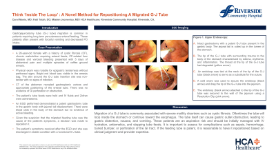
Figure: Figure 1. Upper Endoscopy: A. Intact gastrostomy with a patent G-J tube present in the gastric body. The jejunal tail is coiled up in the lumen of the stomach. B. The tip of the G-J tube with surrounding trauma to the body of the stomach, characterized by edema, erythema, and inflammation. The thread at the tip of the G-J tube had degraded (yellow arrow). C. An endoloop was tied at the neck of the tip of the G-J tube (black arrow) to serve as a substitute for the suture. D. A cold snare was used to secure the endoloop (black arrow) and drag the tip of the G-J tube into the jejunum. E. The endoloop (black arrow) attached to the tip of the G-J tube was secured to the wall of the jejunum using a Resolution Clip (pink arrow).
Disclosures:
Carol Monis indicated no relevant financial relationships.
Fadi Totah indicated no relevant financial relationships.
Mladen Jecmenica indicated no relevant financial relationships.
Carol Monis, MD1, Fadi Totah, DO1, Mladen Jecmenica, MD2. B0294 - Think "Inside the Loop:" A Novel Method for Repositioning a Migrated G-J Tube, ACG 2022 Annual Scientific Meeting Abstracts. Charlotte, NC: American College of Gastroenterology.
