Back


Poster Session E - Tuesday Afternoon
Category: Colon
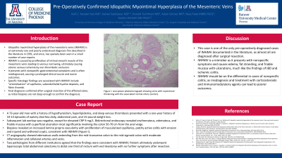
E0132 - Pre-Operatively Confirmed Idiopathic Myointimal Hyperplasia of the Mesenteric Veins
Tuesday, October 25, 2022
3:00 PM – 5:00 PM ET
Location: Crown Ballroom

Has Audio

Kelli C. Kosako Yost, MD
University of Arizona College of Medicine
Phoenix, AZ
Presenting Author(s)
Award: Presidential Poster Award
Kelli C. Kosako Yost, MD1, Yasmin Alishahi, MD, FACG1, Kaivan Salehpour, MD2, Donald Tschirhart, MD1, Adam Gomez, MD3, Neej Patel, MBBS, FACG4
1University of Arizona College of Medicine, Phoenix, AZ; 2Phoenix VA Health Care System, Phoenix, AZ; 3St. Joseph's Hospital and Medical Center, Phoenix, AZ; 4Darmouth Hitchcock Medical Center, Lebanon, NH
Introduction: Idiopathic myointimal hyperplasia of the mesenteric veins (IMHMV) is an extremely rare and poorly understood diagnosis first described in the literature in 1991, and since, has sparsely been seen in a small number of case reports. IMHMV is caused by proliferation of intimal smooth muscle of the mesenteric veins leading to venous narrowing, ultimately causing colonic venous ischemia by non-thrombotic occlusion. It presents with nonspecific gastrointestinal complaints and is often misdiagnosed, causing a prolonged clinical course and worse outcomes. Specific histologic findings are associated with IMHMV include “arteriolization” of capillaries, subendothelial hyaline deposits, and fibrin thrombi. Final diagnosis confirmed after surgical resection of the affected colon, as initial biopsies are not deep enough to confirm the diagnosis.
Case Description/Methods: A 73-year-old man with a history of hypothyroidism, hyperlipidemia, and deep venous thrombosis presented with a one-year history of 10-14 episodes of watery diarrhea daily, abdominal pain, and 15-pound weight loss. Subsequent lab workup was negative, except for elevated CRP 9 mg/L. Bidirectional endoscopy revealed erythematous, edematous, and friable mucosa with superficial ulceration most significantly involving the colon 50-70 cm from the anal verge. Biopsies revealed an increased lamina propria vascularity with proliferation of muscularized capillaries, patchy active colitis with erosion and injured and withered crypts, consistent with IMHMV. CT angiography showed edematous walls extending from the mid-transverse colon to the mid-sigmoid colon with moderate inflammation and collateral arteries and veins. Two pathologists from different institutions agreed that the findings were consistent with IMHMV. Patient ultimately underwent laparoscopic total abdominal colectomy to distal one third of rectum with end ileostomy with no further symptoms after resection.
Discussion: This case is one of the only pre-operatively diagnosed cases of IMHMV documented in the literature, as almost all are diagnosed after surgical resection. IMHMV is a mimicker as it presents with nonspecific symptoms and causes edema, fat stranding, and friable mucosa with ulcerations, much like the findings of IBD and ischemic colitis. IMHMV should be on the differential in cases of nonspecific colitis, as misdiagnosis and treatment with corticosteroids and immunomodulatory agents can lead to poorer outcomes.

Disclosures:
Kelli C. Kosako Yost, MD1, Yasmin Alishahi, MD, FACG1, Kaivan Salehpour, MD2, Donald Tschirhart, MD1, Adam Gomez, MD3, Neej Patel, MBBS, FACG4. E0132 - Pre-Operatively Confirmed Idiopathic Myointimal Hyperplasia of the Mesenteric Veins, ACG 2022 Annual Scientific Meeting Abstracts. Charlotte, NC: American College of Gastroenterology.
Kelli C. Kosako Yost, MD1, Yasmin Alishahi, MD, FACG1, Kaivan Salehpour, MD2, Donald Tschirhart, MD1, Adam Gomez, MD3, Neej Patel, MBBS, FACG4
1University of Arizona College of Medicine, Phoenix, AZ; 2Phoenix VA Health Care System, Phoenix, AZ; 3St. Joseph's Hospital and Medical Center, Phoenix, AZ; 4Darmouth Hitchcock Medical Center, Lebanon, NH
Introduction: Idiopathic myointimal hyperplasia of the mesenteric veins (IMHMV) is an extremely rare and poorly understood diagnosis first described in the literature in 1991, and since, has sparsely been seen in a small number of case reports. IMHMV is caused by proliferation of intimal smooth muscle of the mesenteric veins leading to venous narrowing, ultimately causing colonic venous ischemia by non-thrombotic occlusion. It presents with nonspecific gastrointestinal complaints and is often misdiagnosed, causing a prolonged clinical course and worse outcomes. Specific histologic findings are associated with IMHMV include “arteriolization” of capillaries, subendothelial hyaline deposits, and fibrin thrombi. Final diagnosis confirmed after surgical resection of the affected colon, as initial biopsies are not deep enough to confirm the diagnosis.
Case Description/Methods: A 73-year-old man with a history of hypothyroidism, hyperlipidemia, and deep venous thrombosis presented with a one-year history of 10-14 episodes of watery diarrhea daily, abdominal pain, and 15-pound weight loss. Subsequent lab workup was negative, except for elevated CRP 9 mg/L. Bidirectional endoscopy revealed erythematous, edematous, and friable mucosa with superficial ulceration most significantly involving the colon 50-70 cm from the anal verge. Biopsies revealed an increased lamina propria vascularity with proliferation of muscularized capillaries, patchy active colitis with erosion and injured and withered crypts, consistent with IMHMV. CT angiography showed edematous walls extending from the mid-transverse colon to the mid-sigmoid colon with moderate inflammation and collateral arteries and veins. Two pathologists from different institutions agreed that the findings were consistent with IMHMV. Patient ultimately underwent laparoscopic total abdominal colectomy to distal one third of rectum with end ileostomy with no further symptoms after resection.
Discussion: This case is one of the only pre-operatively diagnosed cases of IMHMV documented in the literature, as almost all are diagnosed after surgical resection. IMHMV is a mimicker as it presents with nonspecific symptoms and causes edema, fat stranding, and friable mucosa with ulcerations, much like the findings of IBD and ischemic colitis. IMHMV should be on the differential in cases of nonspecific colitis, as misdiagnosis and treatment with corticosteroids and immunomodulatory agents can lead to poorer outcomes.

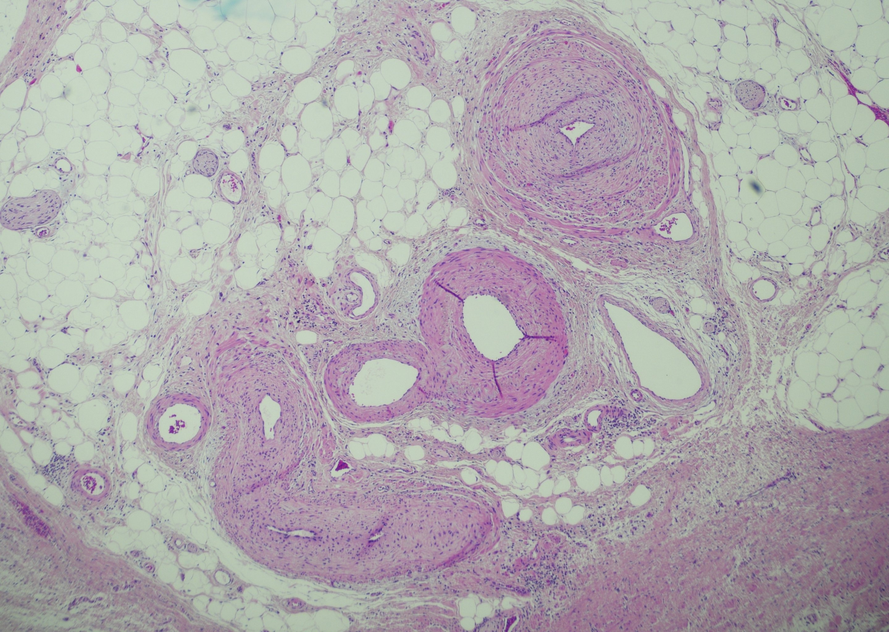
Figure: Low-power photomicrograph showing veins with myointimal thickening with the associated normal artery (center).
Disclosures:
Kelli Kosako Yost indicated no relevant financial relationships.
Yasmin Alishahi indicated no relevant financial relationships.
Kaivan Salehpour indicated no relevant financial relationships.
Donald Tschirhart indicated no relevant financial relationships.
Adam Gomez indicated no relevant financial relationships.
Neej Patel indicated no relevant financial relationships.
Kelli C. Kosako Yost, MD1, Yasmin Alishahi, MD, FACG1, Kaivan Salehpour, MD2, Donald Tschirhart, MD1, Adam Gomez, MD3, Neej Patel, MBBS, FACG4. E0132 - Pre-Operatively Confirmed Idiopathic Myointimal Hyperplasia of the Mesenteric Veins, ACG 2022 Annual Scientific Meeting Abstracts. Charlotte, NC: American College of Gastroenterology.

