Back


Poster Session C - Monday Afternoon
Category: Colon
C0162 - A Rare Case of Symptomatic Colonic Lymphangioma
Monday, October 24, 2022
3:00 PM – 5:00 PM ET
Location: Crown Ballroom

Has Audio

Karla M. Ramos-Feliciano, MD
St. Luke's Hospital - Bethlehem Campus
Allentown, PA
Presenting Author(s)
Karla M. Ramos-Feliciano, MD1, Brian Kim, DO2, Yecheskel Schneider, MD2, Samantha Rollins, DO3
1St. Luke's Hospital - Bethlehem Campus, Allentown, PA; 2St. Luke's University Health Network, Bethlehem, PA; 3St. Luke's University, Bethlehem, PA
Introduction: Lymphangiomas are benign lymphatic lesions, most frequently identified in the head and neck. Colonic lymphangiomas occur but are rare and represent less than 1% of all lymphatic malformations. These lesions are often asymptomatic and discovered incidentally during routine colonoscopy. We present a case of a symptomatic colonic lymphangioma manifesting as a large mass requiring surgical resection.
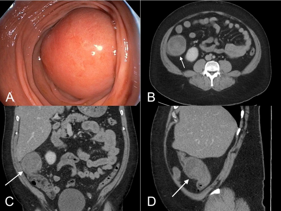
Case Description/Methods: A 42-year-old male presented with the complaint of lower abdominal pain. Associated symptoms included nausea with no vomiting, and he denied family history of gastrointestinal malignancies. At presentation, vital signs were stable and abdominal exam was benign. Laboratory studies were unremarkable, but a Computed Tomography (CT) abdomen/pelvis with contrast revealed a 6.6 x 7 x 6.6 cm colonic mass near the hepatic flexure. Follow up colonoscopy demonstrated a large, subepithelial-appearing mass in the ascending colon protruding more than 90% into the lumen. Retroflexed view revealed 2 x 2 cm ulceration on the proximal aspect of the lesion. Serum tumor markers including CEA, CA 19-9, and CA 125 were all within normal limits and additional CT imaging revealed no metastatic lesions. Biopsies resulted back demonstrating superficial portions of colonic mucosa with mild focal inflammation. However, surgical evaluation was advised given the mass size and the patient thus underwent an elective laparoscopic right hemicolectomy. Tissue samples were sent for evaluation and revealed lymphangiomatosis/lymphatic malformation/cystic lymphangioma centered in the colonic submucosa with extension into the subserosal tissue. No cellular atypia or malignancy was identified, and 24 lymph nodes were benign. These pathology findings were confirmed by expert review at a tertiary center, where it was noted that this case also lacked features of congenital lymphangiectasia. During post-operative follow-up, the patient was reported to be progressing well.
Discussion: Lymphangiomas have an incidence of less than 0.20% and intra-abdominal lymphangiomas represent only a fraction of cases. Although most colonic lymphangiomas are asymptomatic and identified incidentally, they can lead to an array of symptoms and severe complications such as obstruction, intussusception, and perforation requiring surgical intervention if large. Although colonic lymphangiomas are rare, it is important to be aware of their rising incidence to help facilitate accurate and timely diagnosis to prevent complications.

Disclosures:
Karla M. Ramos-Feliciano, MD1, Brian Kim, DO2, Yecheskel Schneider, MD2, Samantha Rollins, DO3. C0162 - A Rare Case of Symptomatic Colonic Lymphangioma, ACG 2022 Annual Scientific Meeting Abstracts. Charlotte, NC: American College of Gastroenterology.
1St. Luke's Hospital - Bethlehem Campus, Allentown, PA; 2St. Luke's University Health Network, Bethlehem, PA; 3St. Luke's University, Bethlehem, PA
Introduction: Lymphangiomas are benign lymphatic lesions, most frequently identified in the head and neck. Colonic lymphangiomas occur but are rare and represent less than 1% of all lymphatic malformations. These lesions are often asymptomatic and discovered incidentally during routine colonoscopy. We present a case of a symptomatic colonic lymphangioma manifesting as a large mass requiring surgical resection.
Case Description/Methods: A 42-year-old male presented with the complaint of lower abdominal pain. Associated symptoms included nausea with no vomiting, and he denied family history of gastrointestinal malignancies. At presentation, vital signs were stable and abdominal exam was benign. Laboratory studies were unremarkable, but a Computed Tomography (CT) abdomen/pelvis with contrast revealed a 6.6 x 7 x 6.6 cm colonic mass near the hepatic flexure. Follow up colonoscopy demonstrated a large, subepithelial-appearing mass in the ascending colon protruding more than 90% into the lumen. Retroflexed view revealed 2 x 2 cm ulceration on the proximal aspect of the lesion. Serum tumor markers including CEA, CA 19-9, and CA 125 were all within normal limits and additional CT imaging revealed no metastatic lesions. Biopsies resulted back demonstrating superficial portions of colonic mucosa with mild focal inflammation. However, surgical evaluation was advised given the mass size and the patient thus underwent an elective laparoscopic right hemicolectomy. Tissue samples were sent for evaluation and revealed lymphangiomatosis/lymphatic malformation/cystic lymphangioma centered in the colonic submucosa with extension into the subserosal tissue. No cellular atypia or malignancy was identified, and 24 lymph nodes were benign. These pathology findings were confirmed by expert review at a tertiary center, where it was noted that this case also lacked features of congenital lymphangiectasia. During post-operative follow-up, the patient was reported to be progressing well.
Discussion: Lymphangiomas have an incidence of less than 0.20% and intra-abdominal lymphangiomas represent only a fraction of cases. Although most colonic lymphangiomas are asymptomatic and identified incidentally, they can lead to an array of symptoms and severe complications such as obstruction, intussusception, and perforation requiring surgical intervention if large. Although colonic lymphangiomas are rare, it is important to be aware of their rising incidence to help facilitate accurate and timely diagnosis to prevent complications.

Figure: Figure A. Colonoscopy. Submucosal mass in the ascending colon involving more than 90% of the circumference.
Figure B. CT axial image. Mass in the ascending colon/hepatic flexure.
Figure C. CT coronal image. Mass in the ascending colon/hepatic flexure.
Figure D. CT sagittal image. Mass in the ascending colon/hepatic flexure.
Figure B. CT axial image. Mass in the ascending colon/hepatic flexure.
Figure C. CT coronal image. Mass in the ascending colon/hepatic flexure.
Figure D. CT sagittal image. Mass in the ascending colon/hepatic flexure.
Disclosures:
Karla Ramos-Feliciano indicated no relevant financial relationships.
Brian Kim indicated no relevant financial relationships.
Yecheskel Schneider indicated no relevant financial relationships.
Samantha Rollins indicated no relevant financial relationships.
Karla M. Ramos-Feliciano, MD1, Brian Kim, DO2, Yecheskel Schneider, MD2, Samantha Rollins, DO3. C0162 - A Rare Case of Symptomatic Colonic Lymphangioma, ACG 2022 Annual Scientific Meeting Abstracts. Charlotte, NC: American College of Gastroenterology.
