Back


Poster Session A - Sunday Afternoon
Category: Interventional Endoscopy
A0445 - A Rare Case of a Esophagomediastinal Fistula Closed Endoscopically in a Young Patient With Tuberculosis and HIV
Sunday, October 23, 2022
5:00 PM – 7:00 PM ET
Location: Crown Ballroom

Has Audio

Vincent Wong, MD
Rutgers New Jersey Medical School
Newark, NJ
Presenting Author(s)
Vincent Wong, MD, Ahmed Ahmed, DO, Anjella Manoharan, MS, MD, Weizheng Wang, MD
Rutgers New Jersey Medical School, Newark, NJ
Introduction: Tuberculosis (TB) is usually a primary lung disease affecting approximately 10 million people worldwide. Extrapulmonary manifestations can involve the lymph nodes, vertebrae, and gastrointestinal tract. In immunocompromised patients, TB can manifest in the esophagus as fistulas with the trachea and bronchus and rarely with the mediastinum, which can lead to increased morbidity and mortality.
Case Description/Methods: We report a 29-year-old male with human immunodeficiency virus (HIV) and tuberculosis, diagnosed recently by mediastinal biopsy, who presented with dysphagia. His symptoms started one month ago with difficulty swallowing solids and thin liquids, causing him to have pleuritic chest pain, cough, and weight loss.
His vitals and basic labs were unremarkable. He is cachetic appearing but in no acute distress with a visible healed scar below the sixth rib. Lung exam had diminished breath sounds in the right lower lung field. CT chest showed trace extraluminal contrast to the right of the esophagus with a small focus of gas within the mediastinum concerning for a esophagomediastinal fistula. Esophagogastroduodenoscopy (EGD) was done that showed esophagitis without bleeding and a fistula in the middle third of the esophagus. Six clips were placed to close the defect and follow-up gastrograffin study did not show contrast extravasation. Our patient received a percutaneous gastrostomy tube for nutrition and fistula healing.
On follow-up, the patient’s symptoms continue to improve without signs of aspiration on chest x-ray.
Discussion: Esophagomediastinal fistulas are rare in patients with tuberculosis. They occur in about 17.6% of patients with TB and 35% of patients with concomitant HIV. Fistula tracts develop spontaneously from ruptured caseating lymph nodes that erode into adjacent organs or from iatrogenic procedures such as biopsies. Patients can present with a cough after eating leading to long term malnutrition and weight loss. The diagnosis can be made with an esophagram, CT, or EGD. It is treated by closing the defect, controlling the infection, and optimizing nutrition. Fistulas are typically closed surgically but they can now be closed endoscopically using clips, stents, and stitches. We were able to demonstrate the first use of through the scope endoclips to successfully close a TB esophagomediastinal fistula. We propose that this method be considered the gold standard because it is low risk, minimally invasive, low-cost, and effective.

Disclosures:
Vincent Wong, MD, Ahmed Ahmed, DO, Anjella Manoharan, MS, MD, Weizheng Wang, MD. A0445 - A Rare Case of a Esophagomediastinal Fistula Closed Endoscopically in a Young Patient With Tuberculosis and HIV, ACG 2022 Annual Scientific Meeting Abstracts. Charlotte, NC: American College of Gastroenterology.
Rutgers New Jersey Medical School, Newark, NJ
Introduction: Tuberculosis (TB) is usually a primary lung disease affecting approximately 10 million people worldwide. Extrapulmonary manifestations can involve the lymph nodes, vertebrae, and gastrointestinal tract. In immunocompromised patients, TB can manifest in the esophagus as fistulas with the trachea and bronchus and rarely with the mediastinum, which can lead to increased morbidity and mortality.
Case Description/Methods: We report a 29-year-old male with human immunodeficiency virus (HIV) and tuberculosis, diagnosed recently by mediastinal biopsy, who presented with dysphagia. His symptoms started one month ago with difficulty swallowing solids and thin liquids, causing him to have pleuritic chest pain, cough, and weight loss.
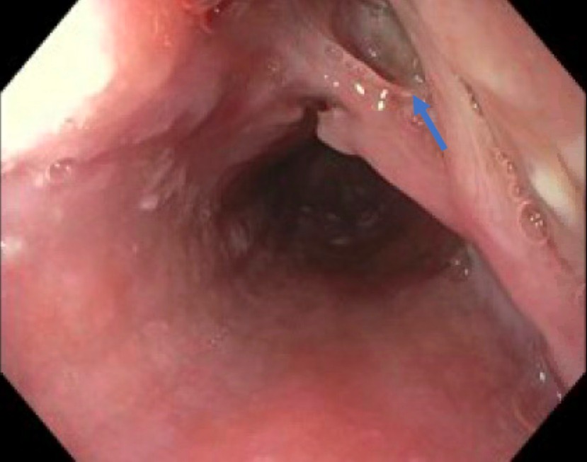
His vitals and basic labs were unremarkable. He is cachetic appearing but in no acute distress with a visible healed scar below the sixth rib. Lung exam had diminished breath sounds in the right lower lung field. CT chest showed trace extraluminal contrast to the right of the esophagus with a small focus of gas within the mediastinum concerning for a esophagomediastinal fistula. Esophagogastroduodenoscopy (EGD) was done that showed esophagitis without bleeding and a fistula in the middle third of the esophagus. Six clips were placed to close the defect and follow-up gastrograffin study did not show contrast extravasation. Our patient received a percutaneous gastrostomy tube for nutrition and fistula healing.
On follow-up, the patient’s symptoms continue to improve without signs of aspiration on chest x-ray.
Discussion: Esophagomediastinal fistulas are rare in patients with tuberculosis. They occur in about 17.6% of patients with TB and 35% of patients with concomitant HIV. Fistula tracts develop spontaneously from ruptured caseating lymph nodes that erode into adjacent organs or from iatrogenic procedures such as biopsies. Patients can present with a cough after eating leading to long term malnutrition and weight loss. The diagnosis can be made with an esophagram, CT, or EGD. It is treated by closing the defect, controlling the infection, and optimizing nutrition. Fistulas are typically closed surgically but they can now be closed endoscopically using clips, stents, and stitches. We were able to demonstrate the first use of through the scope endoclips to successfully close a TB esophagomediastinal fistula. We propose that this method be considered the gold standard because it is low risk, minimally invasive, low-cost, and effective.

Figure: Esophagomediastinal fistula (blue arrow) seen in the middle third of the esophagus on esophagogastroduodenoscopy (EGD).
Disclosures:
Vincent Wong indicated no relevant financial relationships.
Ahmed Ahmed indicated no relevant financial relationships.
Anjella Manoharan indicated no relevant financial relationships.
Weizheng Wang indicated no relevant financial relationships.
Vincent Wong, MD, Ahmed Ahmed, DO, Anjella Manoharan, MS, MD, Weizheng Wang, MD. A0445 - A Rare Case of a Esophagomediastinal Fistula Closed Endoscopically in a Young Patient With Tuberculosis and HIV, ACG 2022 Annual Scientific Meeting Abstracts. Charlotte, NC: American College of Gastroenterology.
