Back


Poster Session C - Monday Afternoon
Category: Stomach
C0724 - A Rare Case of Extramedullary Gastric Plasmacytoma Presenting With Pseudoachalasia
Monday, October 24, 2022
3:00 PM – 5:00 PM ET
Location: Crown Ballroom

Has Audio

Sadaf Afraz, DO
Cleveland Clinic Florida
Weston, Florida
Presenting Author(s)
Sadaf Afraz, DO1, Kanwarpreet Tandon, MD1, Ashraf Almomani, MD2, Adalberto Gonzalez, MD1, Asad ur Rahman, MD1, Tolga Erim, DO1, Julia Diacovo, MD1, Fernando Castro-Pavia, MD1
1Cleveland Clinic Florida, Weston, FL; 2Cleveland Clinic Foundation, Cleveland, OH
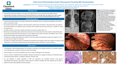
Introduction: Extramedullary plasmacytoma (EMP) is a rare plasma cell neoplasm that can occur in association with multiple myeloma. Gastrointestinal involvement is extremely rare and accounts for less than 5% of all EMP cases. We present here a case of multiple myeloma initially presenting with pseudo-achalasia due to EMP of the stomach involving the gastroesophageal junction (GEJ).
Case Description/Methods: A 65-year-old female presented to the emergency department for symptomatic anemia, and progressive dysphagia to both solid and liquids in the past three months. She reported associated 20 lbs. weight loss. She denied melena, abdominal pain and heartburn. Past medical history was notable for chronic kidney disease and iron deficiency anemia. Physical exam was unremarkable. Laboratory workup revealed microcytic anemia with Hg of 8.1, platelet 167, calcium 8.8, creatinine 2.54. The patient underwent a timed barium swallow with findings concerning for achalasia (Image 1). MRI abdomen showed diffuse gastric wall thickening with extension into the GEJ (Image 1). EGD evaluation revealed friable gastric mucosa with severe thickening of the folds (Image 1). Biopsies were consistent with plasma cell malignancy. Serum immuno-electrophoresis revealed bi-clonal gammopathy of IgA lambda and IgG kappa. The diagnosis of multiple myeloma (MM) was confirmed on bone marrow biopsy with identification of plasma cell neoplasm of 10%. A Congo red stain was negative for amyloidosis. The patient was managed with bortezomib, dexamethasone and daratumumab combined therapy with resolution of symptoms at three months follow up. She achieved complete endoscopic and histological remission of EMP after 13 cycles of treatment.
Discussion: The presentation of MM with GI involvement is extremely rare and typically presents with non-specific symptoms such as abdominal pain, nausea and vomiting, and rarely GI hemorrhage. Pseudo-achalasia is an uncommon presentation and occurs in cases of esophageal or GEJ involvement. There are currently no established treatment guidelines, although most patients are managed with radiation and combination therapy with Bortezomib-based regimens, or autologous transplant in refractory cases. Our case highlights an atypical presentation of EMP with symptomatic and histological resolution using adjuvant chemotherapy/immunotherapy. A high degree of clinical suspicion is essential in facilitating the correct diagnosis given that delay in treatment can lead to rapid progression and poor prognosis.

Disclosures:
Sadaf Afraz, DO1, Kanwarpreet Tandon, MD1, Ashraf Almomani, MD2, Adalberto Gonzalez, MD1, Asad ur Rahman, MD1, Tolga Erim, DO1, Julia Diacovo, MD1, Fernando Castro-Pavia, MD1. C0724 - A Rare Case of Extramedullary Gastric Plasmacytoma Presenting With Pseudoachalasia, ACG 2022 Annual Scientific Meeting Abstracts. Charlotte, NC: American College of Gastroenterology.
1Cleveland Clinic Florida, Weston, FL; 2Cleveland Clinic Foundation, Cleveland, OH
Introduction: Extramedullary plasmacytoma (EMP) is a rare plasma cell neoplasm that can occur in association with multiple myeloma. Gastrointestinal involvement is extremely rare and accounts for less than 5% of all EMP cases. We present here a case of multiple myeloma initially presenting with pseudo-achalasia due to EMP of the stomach involving the gastroesophageal junction (GEJ).
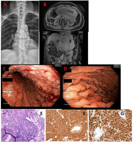
Case Description/Methods: A 65-year-old female presented to the emergency department for symptomatic anemia, and progressive dysphagia to both solid and liquids in the past three months. She reported associated 20 lbs. weight loss. She denied melena, abdominal pain and heartburn. Past medical history was notable for chronic kidney disease and iron deficiency anemia. Physical exam was unremarkable. Laboratory workup revealed microcytic anemia with Hg of 8.1, platelet 167, calcium 8.8, creatinine 2.54. The patient underwent a timed barium swallow with findings concerning for achalasia (Image 1). MRI abdomen showed diffuse gastric wall thickening with extension into the GEJ (Image 1). EGD evaluation revealed friable gastric mucosa with severe thickening of the folds (Image 1). Biopsies were consistent with plasma cell malignancy. Serum immuno-electrophoresis revealed bi-clonal gammopathy of IgA lambda and IgG kappa. The diagnosis of multiple myeloma (MM) was confirmed on bone marrow biopsy with identification of plasma cell neoplasm of 10%. A Congo red stain was negative for amyloidosis. The patient was managed with bortezomib, dexamethasone and daratumumab combined therapy with resolution of symptoms at three months follow up. She achieved complete endoscopic and histological remission of EMP after 13 cycles of treatment.
Discussion: The presentation of MM with GI involvement is extremely rare and typically presents with non-specific symptoms such as abdominal pain, nausea and vomiting, and rarely GI hemorrhage. Pseudo-achalasia is an uncommon presentation and occurs in cases of esophageal or GEJ involvement. There are currently no established treatment guidelines, although most patients are managed with radiation and combination therapy with Bortezomib-based regimens, or autologous transplant in refractory cases. Our case highlights an atypical presentation of EMP with symptomatic and histological resolution using adjuvant chemotherapy/immunotherapy. A high degree of clinical suspicion is essential in facilitating the correct diagnosis given that delay in treatment can lead to rapid progression and poor prognosis.

Figure: Image 1: (A) Timed barium esophagram showing dilated and tortuous esophagus. Beaking of the esophagus at the GE junction region with delayed passage of contrast into the stomach. (B) MRI abdomen with contrast showing very severe diffuse gastric wall thickening. (C)Esophagogastroduodenoscopy at the time of diagnosis showing severe diffuse thickening with friable mucosa from the gastric cardia to the antrum, (D) Improvement post treatment. (F)Antral-type gastric mucosa diffusely infiltrated by a monotonous population of atypical plasma cell infiltrates (Hematoxylin and eosin,100X). (G) Strongly positive CD138. (H) MUM1 (Immunohistochemistry,200X).
Disclosures:
Sadaf Afraz indicated no relevant financial relationships.
Kanwarpreet Tandon indicated no relevant financial relationships.
Ashraf Almomani indicated no relevant financial relationships.
Adalberto Gonzalez indicated no relevant financial relationships.
Asad ur Rahman indicated no relevant financial relationships.
Tolga Erim indicated no relevant financial relationships.
Julia Diacovo indicated no relevant financial relationships.
Fernando Castro-Pavia indicated no relevant financial relationships.
Sadaf Afraz, DO1, Kanwarpreet Tandon, MD1, Ashraf Almomani, MD2, Adalberto Gonzalez, MD1, Asad ur Rahman, MD1, Tolga Erim, DO1, Julia Diacovo, MD1, Fernando Castro-Pavia, MD1. C0724 - A Rare Case of Extramedullary Gastric Plasmacytoma Presenting With Pseudoachalasia, ACG 2022 Annual Scientific Meeting Abstracts. Charlotte, NC: American College of Gastroenterology.
