Back


Poster Session E - Tuesday Afternoon
Category: Colon
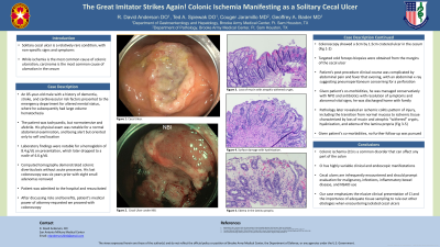
E0155 - The Great Imitator Strikes Again! Colonic Ischemia Manifesting as a Solitary Cecal Ulcer
Tuesday, October 25, 2022
3:00 PM – 5:00 PM ET
Location: Crown Ballroom

Has Audio

R. David Anderson, DO
San Antonio Military Medical Center
Fort Sam Houston, TX
Presenting Author(s)
R. David Anderson, DO, Ted A. Spiewak, DO, Couger Jaramillo, MD, Geoffrey Bader, MD
San Antonio Military Medical Center, Fort Sam Houston, TX
Introduction: Solitary cecal ulcer is a relatively rare condition, with non-specific signs and symptoms. While ischemia is
the most common cause of colonic ulceration, carcinoma is the most common cause of ulceration in the
cecum. We present a unique case of a solitary cecal ulcer secondary to ischemia to highlight the
notoriously variable presentation of colonic ischemia.
Case Description/Methods: An 85-year-old male with a history of dementia, stroke, and cardiovascular risk factors presented to the
emergency department for altered mental status, where he subsequently had large volume
hematochezia. The patient was tachycardic, but normotensive and afebrile. His physical exam was
notable for a normal abdominal examination, and being alert but oriented only to self and location.
Laboratory findings were notable for a hemoglobin of 8.4 g/dL on presentation, which later dropped to a
nadir of 4.6 g/dL. Computed tomography demonstrated colonic diverticulosis without acute processes.
His last colonoscopy was six years prior with eight adenomas. Patient was admitted to the hospital and
resuscitated.
After discussing risks and benefits, patient’s medical power of attorney requested we proceed with
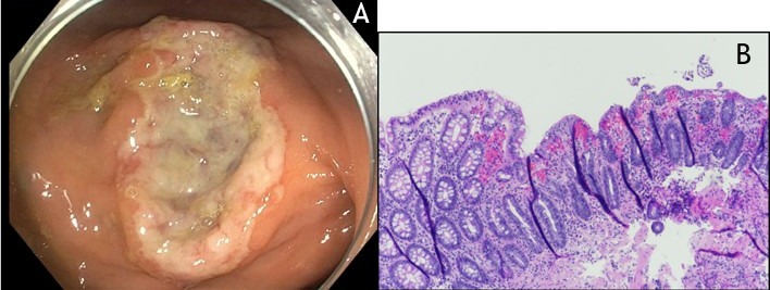
colonoscopy. Colonoscopy showed a 3cm by 1.5cm cratered ulcer in the cecum (Fig 1 A). Targeted cold
forceps biopsies were obtained from the margins of the cecal ulcer. Patient’s clinical course was
complicated by abdominal pain and fever that evening, with an abdominal x-ray suggesting
pneumoperitoneum concerning for a perforation. Given patient’s co-morbidities, he was managed
conservatively with NPO and antibiotics with resolution of symptoms and abnormal vital signs; he was
ultimately discharged home with family. Pathology later revealed an ischemic colitis pattern of injury,
including the transition from normal mucosa to ischemic tissue characterized by loss of mucin and
atrophic “withered” crypts, hyalinization, and edema of the lamina propria (Fig 1 B). Given patient’s co-
morbidities, no further follow up was pursued.
Discussion: Colonic ischemia is a common disorder that can affect any part of the colon, with highly variable clinical
and endoscopic manifestations. Less commonly encountered are isolated cecal ulcers, which should
prompt the evaluation for malignancy, infections, inflammatory bowel disease, or NSAID use. This case
emphasizes the elusive clinical presentation of colonic ischemia and the importance of adequate tissue
sampling to rule out other etiologies when encountering isolated cecal ulcers.

Disclosures:
R. David Anderson, DO, Ted A. Spiewak, DO, Couger Jaramillo, MD, Geoffrey Bader, MD. E0155 - The Great Imitator Strikes Again! Colonic Ischemia Manifesting as a Solitary Cecal Ulcer, ACG 2022 Annual Scientific Meeting Abstracts. Charlotte, NC: American College of Gastroenterology.
San Antonio Military Medical Center, Fort Sam Houston, TX
Introduction: Solitary cecal ulcer is a relatively rare condition, with non-specific signs and symptoms. While ischemia is
the most common cause of colonic ulceration, carcinoma is the most common cause of ulceration in the
cecum. We present a unique case of a solitary cecal ulcer secondary to ischemia to highlight the
notoriously variable presentation of colonic ischemia.
Case Description/Methods: An 85-year-old male with a history of dementia, stroke, and cardiovascular risk factors presented to the
emergency department for altered mental status, where he subsequently had large volume
hematochezia. The patient was tachycardic, but normotensive and afebrile. His physical exam was
notable for a normal abdominal examination, and being alert but oriented only to self and location.
Laboratory findings were notable for a hemoglobin of 8.4 g/dL on presentation, which later dropped to a
nadir of 4.6 g/dL. Computed tomography demonstrated colonic diverticulosis without acute processes.
His last colonoscopy was six years prior with eight adenomas. Patient was admitted to the hospital and
resuscitated.
After discussing risks and benefits, patient’s medical power of attorney requested we proceed with
colonoscopy. Colonoscopy showed a 3cm by 1.5cm cratered ulcer in the cecum (Fig 1 A). Targeted cold
forceps biopsies were obtained from the margins of the cecal ulcer. Patient’s clinical course was
complicated by abdominal pain and fever that evening, with an abdominal x-ray suggesting
pneumoperitoneum concerning for a perforation. Given patient’s co-morbidities, he was managed
conservatively with NPO and antibiotics with resolution of symptoms and abnormal vital signs; he was
ultimately discharged home with family. Pathology later revealed an ischemic colitis pattern of injury,
including the transition from normal mucosa to ischemic tissue characterized by loss of mucin and
atrophic “withered” crypts, hyalinization, and edema of the lamina propria (Fig 1 B). Given patient’s co-
morbidities, no further follow up was pursued.
Discussion: Colonic ischemia is a common disorder that can affect any part of the colon, with highly variable clinical
and endoscopic manifestations. Less commonly encountered are isolated cecal ulcers, which should
prompt the evaluation for malignancy, infections, inflammatory bowel disease, or NSAID use. This case
emphasizes the elusive clinical presentation of colonic ischemia and the importance of adequate tissue
sampling to rule out other etiologies when encountering isolated cecal ulcers.

Figure: Fig 1 A is a 3cm by 1.5cm cratered ulcer in the cecum. Fig 1 B is the histology from biopsies of the edge of the ulcer showing an ischemic colitis pattern of injury, including the transition from normal mucosa to ischemic tissue characterized by loss of mucin and atrophic "withered" crypts, hyalinization, and edema of the lamina propria.
Disclosures:
R. David Anderson indicated no relevant financial relationships.
Ted Spiewak indicated no relevant financial relationships.
Couger Jaramillo indicated no relevant financial relationships.
Geoffrey Bader indicated no relevant financial relationships.
R. David Anderson, DO, Ted A. Spiewak, DO, Couger Jaramillo, MD, Geoffrey Bader, MD. E0155 - The Great Imitator Strikes Again! Colonic Ischemia Manifesting as a Solitary Cecal Ulcer, ACG 2022 Annual Scientific Meeting Abstracts. Charlotte, NC: American College of Gastroenterology.
