Back


Poster Session A - Sunday Afternoon
Category: Pediatrics
A0611 - To Bead or Not to Bead: Use of Ultrasound and a Conservative Approach to Water Bead Ingestion in an Infant
Sunday, October 23, 2022
5:00 PM – 7:00 PM ET
Location: Crown Ballroom

Has Audio

Kacie H. Denton, MD, MPH
East Tennessee State University
Johnson City, TN
Presenting Author(s)
Eliana H. Kim, MD1, Kacie H. Denton, MD, MPH1, Anjali Malkani, MD1, John W. Schweitzer, MD1, Garrett Woodbury, MD2
1East Tennessee State University, Johnson City, TN; 2Empire Radiology, Johnson City, TN
Introduction: Water beads are expanding, non-toxic toys made of water-absorbing polymer. Ingestion of such beads has the potential to cause bowel obstruction due to its ability to continually expand. There are few case reports of ingestion of water beads in infants, however, many required surgical intervention. Here, we describe an infant with water bead ingestions whose symptoms resolved with conservative management, using ultrasound as a supportive imaging modality.
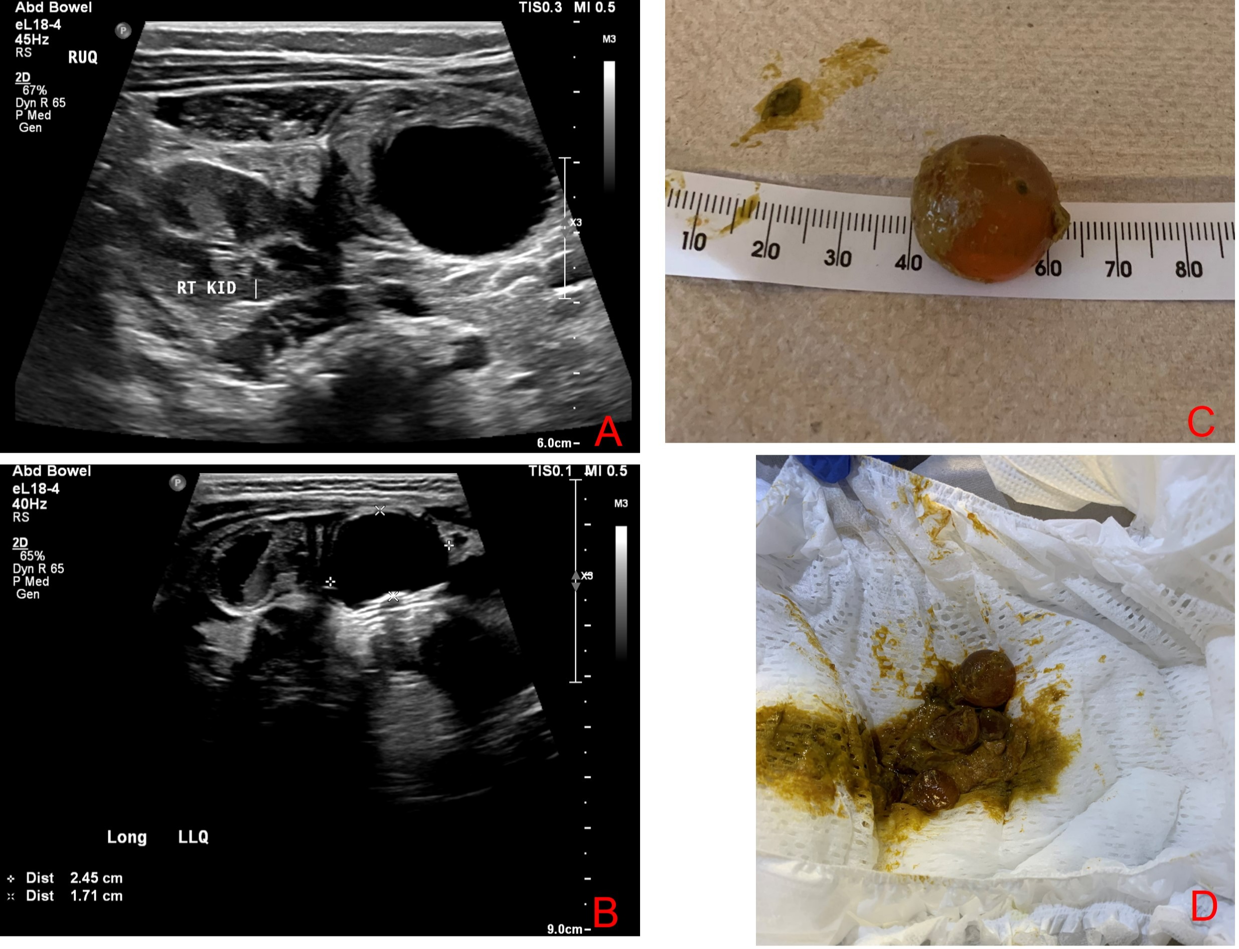
Case Description/Methods: A 7-month-old male presented to the emergency department with gagging and poor intake after playing with water beads approximately 16 hours prior to presentation. Physical exam was normal. No foreign body was noted on nose-to-rectum Xray. Pediatric gastroenterology was consulted for esophagogastroduodenoscopy (EGD). Upon intubation, there was concern for foreign body in the right main bronchus due to decreased breath sounds. No foreign body was visible on either rigid bronchoscopy or subsequent EGD. An ultrasound confirmed a circular anechoic structure in the right upper quadrant, likely the small bowel. He was admitted for observation and made NPO due to the potential for developing bowel obstruction. A second ultrasound 16 hours later revealed an increase in size from 2.2cm to 2.5cm and progression to the left lower quadrant, unclear if located in the colon or small bowel. With a benign abdominal exam, the decision was made to resume breastfeeding. Soon after, he passed two stools containing
multiple fragments of ruptured water beads and one fully intact bead measuring 2 cm. He was discharged home with no further reported problems.
Discussion: The gastrocolic reflex from breastfeeding most likely contributed to our patient’s ability to pass the water bead without further medical or surgical intervention. The use of ultrasound, while helpful, is not a widely recognized modality for detection of ingested water beads because it may not accurately depict the number, size, or location. Clinicians must consider the patient’s clinical presentation, timing post-ingestion, diet, and suspected size and number of water beads in the management. Future considerations in the management include the utilization of osmotic agents, such as gastrografin or polyethylene glycol, to osmotically impact the bead’s ability to enlarge further and thus avoid surgical intervention.

Disclosures:
Eliana H. Kim, MD1, Kacie H. Denton, MD, MPH1, Anjali Malkani, MD1, John W. Schweitzer, MD1, Garrett Woodbury, MD2. A0611 - To Bead or Not to Bead: Use of Ultrasound and a Conservative Approach to Water Bead Ingestion in an Infant, ACG 2022 Annual Scientific Meeting Abstracts. Charlotte, NC: American College of Gastroenterology.
1East Tennessee State University, Johnson City, TN; 2Empire Radiology, Johnson City, TN
Introduction: Water beads are expanding, non-toxic toys made of water-absorbing polymer. Ingestion of such beads has the potential to cause bowel obstruction due to its ability to continually expand. There are few case reports of ingestion of water beads in infants, however, many required surgical intervention. Here, we describe an infant with water bead ingestions whose symptoms resolved with conservative management, using ultrasound as a supportive imaging modality.
Case Description/Methods: A 7-month-old male presented to the emergency department with gagging and poor intake after playing with water beads approximately 16 hours prior to presentation. Physical exam was normal. No foreign body was noted on nose-to-rectum Xray. Pediatric gastroenterology was consulted for esophagogastroduodenoscopy (EGD). Upon intubation, there was concern for foreign body in the right main bronchus due to decreased breath sounds. No foreign body was visible on either rigid bronchoscopy or subsequent EGD. An ultrasound confirmed a circular anechoic structure in the right upper quadrant, likely the small bowel. He was admitted for observation and made NPO due to the potential for developing bowel obstruction. A second ultrasound 16 hours later revealed an increase in size from 2.2cm to 2.5cm and progression to the left lower quadrant, unclear if located in the colon or small bowel. With a benign abdominal exam, the decision was made to resume breastfeeding. Soon after, he passed two stools containing
multiple fragments of ruptured water beads and one fully intact bead measuring 2 cm. He was discharged home with no further reported problems.
Discussion: The gastrocolic reflex from breastfeeding most likely contributed to our patient’s ability to pass the water bead without further medical or surgical intervention. The use of ultrasound, while helpful, is not a widely recognized modality for detection of ingested water beads because it may not accurately depict the number, size, or location. Clinicians must consider the patient’s clinical presentation, timing post-ingestion, diet, and suspected size and number of water beads in the management. Future considerations in the management include the utilization of osmotic agents, such as gastrografin or polyethylene glycol, to osmotically impact the bead’s ability to enlarge further and thus avoid surgical intervention.

Figure: A. Initial ultrasound with circular anechoic structure in right upper quadrant. B Repeat ultrasound with circular anechoic structure now in left lower quadrant, slightly larger in size. C. Intact water bead found in stool. D. Water beads with one intact bead and multiple fragments.
Disclosures:
Eliana Kim indicated no relevant financial relationships.
Kacie Denton indicated no relevant financial relationships.
Anjali Malkani indicated no relevant financial relationships.
John Schweitzer indicated no relevant financial relationships.
Garrett Woodbury indicated no relevant financial relationships.
Eliana H. Kim, MD1, Kacie H. Denton, MD, MPH1, Anjali Malkani, MD1, John W. Schweitzer, MD1, Garrett Woodbury, MD2. A0611 - To Bead or Not to Bead: Use of Ultrasound and a Conservative Approach to Water Bead Ingestion in an Infant, ACG 2022 Annual Scientific Meeting Abstracts. Charlotte, NC: American College of Gastroenterology.
