Back


Poster Session E - Tuesday Afternoon
Category: Endoscopy Video Forum
E0186 - “Into the Folds:” Case Report of a Polypoid Variant of Menetrier’s Disease
Tuesday, October 25, 2022
3:00 PM – 5:00 PM ET
Location: Crown Ballroom

Has Audio

Emily Minor, MD, PhD
University of Virginia
Charlottesville, Virginia
Presenting Author(s)
Emily Minor, MD, PhD1, Mark Radlinski, MD1, Vanessa M. Shami, MD, FACG2
1University of Virginia, Charlottesville, VA; 2University of Virginia Digestive Health Center, Charlottesville, VA
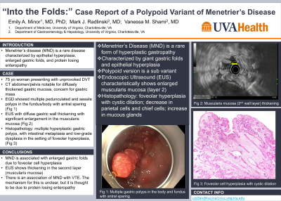
Introduction: Menetrier’s disease (MND) is a rare disease characterized by epithelial hyperplasia, enlarged gastric folds and, protein losing enteropathy. Here we discuss a case of a patient with the polyploid variant of MND who presented with an unprovoked deep vein thrombosis (DVT).
Case Description/Methods: Our patient is a 75-year-old woman who recently developed an unprovoked DVT (placed on rivaroxaban). CT abdomen/pelvis at this time was notable for diffusely thickened gastric mucosa with retroperitoneal soft tissue nodules concerning for metastatic disease. EGD (esophagogastric duodenoscopy) was performed and there was note of “many polypoid lesions” in the fundus and body along with a large, 5-6 cm mass (biopsied). Histopathology showed fragments of gastric non-oxyntic mucosa, foveolar metaplasia, and no overt malignancy. She was referred to us for EUS staging and further management of a presumed gastric malignancy.
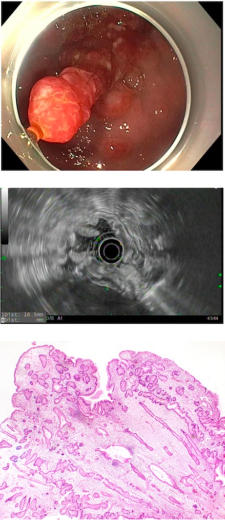
On our examination, EGD revealed multiple pedunculated and sessile polyps in the fundus/body with antral sparing. EUS demonstrated diffuse gastric wall thickening (16.5 mm) with significant enlargement in the second endosonographic layer corresponding to the muscularis mucosae. A large, representative, polyp was removed via snare mucosal resection. Pathology showed multiple hyperplastic gastric polyps, with intestinal metaplasia and low-grade dysplasia in the setting of foveolar hyperplasia, consistent with a polypoid variant of MND.
Discussion: MND is a rare form of hyperplastic gastropathy. Typically, this presents endoscopically with giant gastric folds but there also exists a polypoid variant, as is described with our patient. Diagnosis is made with EGD, EUS, and pathology. Endoscopically, these hyperplastic polyps typically can be found in the fundus and body of the stomach with antral sparing as noted in our patient. Large snare biopsies can be useful in obtaining full thickness epithelial samples. Further confirmation of MND can be made with typical EUS findings including diffuse gastric wall thickening, particularly in the muscularis mucosae (2nd endosonographic layer, hypoechoic). Pathology from gastric resection usually demonstrates foveal hyperplasia with cystic dilation and an increase in mucous glands.
Prior case reports have also noted unprovoked DVT as a presenting symptom of MND. The mechanism for this is unclear but it is thought that protein losing enteropathy associated with MND predisposes to a thrombophilic state with loss of antithrombin III and protein C and S.

Disclosures:
Emily Minor, MD, PhD1, Mark Radlinski, MD1, Vanessa M. Shami, MD, FACG2. E0186 - “Into the Folds:” Case Report of a Polypoid Variant of Menetrier’s Disease, ACG 2022 Annual Scientific Meeting Abstracts. Charlotte, NC: American College of Gastroenterology.
1University of Virginia, Charlottesville, VA; 2University of Virginia Digestive Health Center, Charlottesville, VA
Introduction: Menetrier’s disease (MND) is a rare disease characterized by epithelial hyperplasia, enlarged gastric folds and, protein losing enteropathy. Here we discuss a case of a patient with the polyploid variant of MND who presented with an unprovoked deep vein thrombosis (DVT).
Case Description/Methods: Our patient is a 75-year-old woman who recently developed an unprovoked DVT (placed on rivaroxaban). CT abdomen/pelvis at this time was notable for diffusely thickened gastric mucosa with retroperitoneal soft tissue nodules concerning for metastatic disease. EGD (esophagogastric duodenoscopy) was performed and there was note of “many polypoid lesions” in the fundus and body along with a large, 5-6 cm mass (biopsied). Histopathology showed fragments of gastric non-oxyntic mucosa, foveolar metaplasia, and no overt malignancy. She was referred to us for EUS staging and further management of a presumed gastric malignancy.
On our examination, EGD revealed multiple pedunculated and sessile polyps in the fundus/body with antral sparing. EUS demonstrated diffuse gastric wall thickening (16.5 mm) with significant enlargement in the second endosonographic layer corresponding to the muscularis mucosae. A large, representative, polyp was removed via snare mucosal resection. Pathology showed multiple hyperplastic gastric polyps, with intestinal metaplasia and low-grade dysplasia in the setting of foveolar hyperplasia, consistent with a polypoid variant of MND.
Discussion: MND is a rare form of hyperplastic gastropathy. Typically, this presents endoscopically with giant gastric folds but there also exists a polypoid variant, as is described with our patient. Diagnosis is made with EGD, EUS, and pathology. Endoscopically, these hyperplastic polyps typically can be found in the fundus and body of the stomach with antral sparing as noted in our patient. Large snare biopsies can be useful in obtaining full thickness epithelial samples. Further confirmation of MND can be made with typical EUS findings including diffuse gastric wall thickening, particularly in the muscularis mucosae (2nd endosonographic layer, hypoechoic). Pathology from gastric resection usually demonstrates foveal hyperplasia with cystic dilation and an increase in mucous glands.
Prior case reports have also noted unprovoked DVT as a presenting symptom of MND. The mechanism for this is unclear but it is thought that protein losing enteropathy associated with MND predisposes to a thrombophilic state with loss of antithrombin III and protein C and S.

Figure: Figure 1: Multiple gastric polyps in the body and fundus with antral sparing.
Figure 2: Muscularis mucosa thickening on EUS. Gastric wall measures 16.5 mm.
Figure 3: Foveolar Cell Hyperplasia on histopathology.
Figure 2: Muscularis mucosa thickening on EUS. Gastric wall measures 16.5 mm.
Figure 3: Foveolar Cell Hyperplasia on histopathology.
Disclosures:
Emily Minor indicated no relevant financial relationships.
Mark Radlinski indicated no relevant financial relationships.
Vanessa Shami: Boston Scientific – Giving a lecture funded by Boston Scientific at ACG interventional fellows course. cook medical – Consultant. olympus – Consultant.
Emily Minor, MD, PhD1, Mark Radlinski, MD1, Vanessa M. Shami, MD, FACG2. E0186 - “Into the Folds:” Case Report of a Polypoid Variant of Menetrier’s Disease, ACG 2022 Annual Scientific Meeting Abstracts. Charlotte, NC: American College of Gastroenterology.
