Back


Poster Session D - Tuesday Morning
Category: Small Intestine
D0680 - Rumble in the Tummy: Atypical Location of Abdominal Thrombosis Post-COVID
Tuesday, October 25, 2022
10:00 AM – 12:00 PM ET
Location: Crown Ballroom

Has Audio

Idrees Suliman, MD
Mountain Vista Medical Center
Mesa, AZ
Presenting Author(s)
Preeyanka Sundar, MD1, Suma Harsha Kosuru, MBBS1, Idrees Suliman, MD2, Sara Ancello, DO1
1Midwestern University, Mesa, AZ; 2Mountain Vista Medical Center, Mesa, AZ
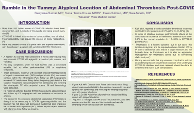
Introduction: More than 532 million cases of COVID-19 infection have been documented, and hundreds of thousands are being added every day. COVID-19 is linked to a number of co-morbidities, one of which, hypercoagulability, has piqued the interest of many researchers. Here, we present a case of a portal vein and superior mesenteric vein thrombosis in a patient with subclinical COVID-19 infection.
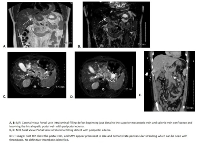
Case Description/Methods: A healthy 30-year-old male presented 3 weeks after having mild, asymptomatic COVID with epigastric abdominal pain, nausea, and vomiting. His only complaints when he had COVID was a decreased appetite, but no other gastrointestinal (GI) or respiratory symptoms. Upon admission, initial CT with contrast showed thrombophlebitis of superior mesenteric vein (SMV) and portal vein (PV), decreased contrast within the intrahepatic PVs; follow up MR Angiography revealed PV intraluminal filling defect beginning just distal to the superior mesenteric vein and splenic vein confluence and involving the intrahepatic PV with periportal edema. He denied any prior clots nor family history of clotting disorder. GI and hematology were consulted. He received catheter-directed tPA for 2 days due to abdominal pain and follow up CT angiogram with no evidence of occlusion in arteries. The cause for his extensive abdominal vasculature thrombosis is thought to be secondary to COVID hypercoagulability, and the location has not been well delineated. Abdominal pain improved, he continued anticoagulation (AC), did not require repeat sessions with plans for close follow up imaging.
Discussion: Klok et al. reported a crude cumulative thrombosis incidence in COVID19 ICU patients is of 57% (95% CI 47–67%). In terms of intestinal damage, prothrombotic effects of the virus rises the incidence of mesenteric ischemia from 0.09–0.2% in the normal population to 1.9–3.8% in COVID-19 patients. Thrombosis is a known outcome, but in our case, this location is atypical, and he required catheter directed tPA by IR due to abdominal pain, that is a large measure and not typically done for thrombosis so it is also an aggressive management for thrombosis (done due to unrelenting abdominal pain). Hereby, we conclude that any vascular complication without an underlying reason should raise suspicion of an underlying COVID-19 infection, and more research should be done to determine the risk of such complications.

Disclosures:
Preeyanka Sundar, MD1, Suma Harsha Kosuru, MBBS1, Idrees Suliman, MD2, Sara Ancello, DO1. D0680 - Rumble in the Tummy: Atypical Location of Abdominal Thrombosis Post-COVID, ACG 2022 Annual Scientific Meeting Abstracts. Charlotte, NC: American College of Gastroenterology.
1Midwestern University, Mesa, AZ; 2Mountain Vista Medical Center, Mesa, AZ
Introduction: More than 532 million cases of COVID-19 infection have been documented, and hundreds of thousands are being added every day. COVID-19 is linked to a number of co-morbidities, one of which, hypercoagulability, has piqued the interest of many researchers. Here, we present a case of a portal vein and superior mesenteric vein thrombosis in a patient with subclinical COVID-19 infection.
Case Description/Methods: A healthy 30-year-old male presented 3 weeks after having mild, asymptomatic COVID with epigastric abdominal pain, nausea, and vomiting. His only complaints when he had COVID was a decreased appetite, but no other gastrointestinal (GI) or respiratory symptoms. Upon admission, initial CT with contrast showed thrombophlebitis of superior mesenteric vein (SMV) and portal vein (PV), decreased contrast within the intrahepatic PVs; follow up MR Angiography revealed PV intraluminal filling defect beginning just distal to the superior mesenteric vein and splenic vein confluence and involving the intrahepatic PV with periportal edema. He denied any prior clots nor family history of clotting disorder. GI and hematology were consulted. He received catheter-directed tPA for 2 days due to abdominal pain and follow up CT angiogram with no evidence of occlusion in arteries. The cause for his extensive abdominal vasculature thrombosis is thought to be secondary to COVID hypercoagulability, and the location has not been well delineated. Abdominal pain improved, he continued anticoagulation (AC), did not require repeat sessions with plans for close follow up imaging.
Discussion: Klok et al. reported a crude cumulative thrombosis incidence in COVID19 ICU patients is of 57% (95% CI 47–67%). In terms of intestinal damage, prothrombotic effects of the virus rises the incidence of mesenteric ischemia from 0.09–0.2% in the normal population to 1.9–3.8% in COVID-19 patients. Thrombosis is a known outcome, but in our case, this location is atypical, and he required catheter directed tPA by IR due to abdominal pain, that is a large measure and not typically done for thrombosis so it is also an aggressive management for thrombosis (done due to unrelenting abdominal pain). Hereby, we conclude that any vascular complication without an underlying reason should raise suspicion of an underlying COVID-19 infection, and more research should be done to determine the risk of such complications.

Figure: Figure A,B: MRI Coronal view. Portal vein intraluminal filling defect beginning just distal to the
superior mesenteric vein and splenic vein confluence and involving the intrahepatic
portal vein with periportal edema
Figure C,D: MRI Axial View of portal vein intraluminal filling defect with periportal edema
Figure E: CT images post tPA show The portal vein, and SMV appear prominent in size and demonstrate
perivascular stranding which can be seen with thrombosis.
Disclosures:
Preeyanka Sundar indicated no relevant financial relationships.
Suma Harsha Kosuru indicated no relevant financial relationships.
Idrees Suliman indicated no relevant financial relationships.
Sara Ancello indicated no relevant financial relationships.
Preeyanka Sundar, MD1, Suma Harsha Kosuru, MBBS1, Idrees Suliman, MD2, Sara Ancello, DO1. D0680 - Rumble in the Tummy: Atypical Location of Abdominal Thrombosis Post-COVID, ACG 2022 Annual Scientific Meeting Abstracts. Charlotte, NC: American College of Gastroenterology.

