Back


Poster Session E - Tuesday Afternoon
Category: Colon
E0124 - Unusual Presentation of Marginal Zone Low Grade B Cell Lymphoma With Extranodal Involvement as a Large Rectal Mass
Tuesday, October 25, 2022
3:00 PM – 5:00 PM ET
Location: Crown Ballroom

Has Audio
.jpg)
Neha Khan, MD
St. Josephs Medical Center, Creighton University
Phoenix, AZ
Presenting Author(s)
Neha Khan, MD1, Kayvon Sotoudeh, MD1, Aida Rezaie, MD2, Hadiatou Barry, MD, MPH3, Brett Hughes, MD1, Savio Redddymasu, MD1
1St. Josephs Medical Center, Creighton University, Phoenix, AZ; 2Creighton University, Phoenix, AZ; 3Creighton University/St. Joseph Hospital and Medical Center, Phoenix, AZ
Introduction: Marginal zone low grade B cell lymphoma represents about 7-8% of all non-Hodgkins lymphomas A subtype of marginal zone B cell lymphoma known as extra-nodal marginal zone lymphoma (MZL) of mucosa-associated lymphoid tissue (MALT Lymphoma) commonly occurs in the gastrointestinal tract, with more than half of all cases occurring in the stomach. While the stomach is the most common location, up to 9% occur in the small intestine and rectal involvement is exceedingly rare. MALT-lymphoma typically takes an indolent course with no B symptoms, whereas patients with nodal MZL present with a more aggressive disease course. Given that MZL rarely presents in the rectum, there is no consensus on treatment guidelines. There is also minimal literature on treatment options for a MZL rectal mass of a substantial size, which provided a unique challenge for this case. We present an atypical presentation of a large (greater than 10cm) marginal zone lymphoma involving the rectum who presented with alternating constipation and incontinence.
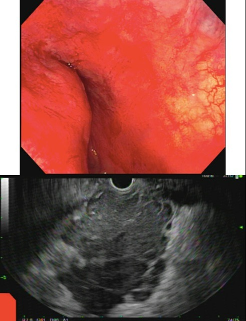
Case Description/Methods: A 73 year old female with a known history of untreated non-Hodgkins lymphoma and prior splenectomy presented with a 3 month history of constipation and fecal incontinence. Computed tomography (CT) scan of the abdomen and pelvis with contrast performed at an outside facility showed an 8 cm abnormal soft tissue mass in the pre-sacral space, causing bowel obstruction at the level of the rectum and anus. The patient subsequently underwent magnetic resonance imaging of the pelvis, which showed a large lobulated mass in the pre-sacral space with suspicion for submucosal origin. A lower endoscopic ultrasound (EUS) with fine needle biopsy was performed which showed a greater than 10 cm large lobulated hypoechoic mass in continuity with the posterior wall of the mid and distal rectum, extending into the anal sphincter. Pathology revealed a low grade B-cell lymphoma with a pattern consistent with marginal zone lymphoma. A bone marrow biopsy was consistent with marginal zone lymphoma. The patient was ultimately discharged with outpatient oncology follow up to start systemic chemotherapy
Discussion: Marginal zone B cell lymphoma presenting as a large rectal mass is a very unique presentation of this neoplastic process. This finding has been reported infrequently in the literature. In this case, lower endoscopic ultrasound (EUS) with FNB was able to be successfully utilized to make the diagnosis and expedite this patient’s treatment course.

Disclosures:
Neha Khan, MD1, Kayvon Sotoudeh, MD1, Aida Rezaie, MD2, Hadiatou Barry, MD, MPH3, Brett Hughes, MD1, Savio Redddymasu, MD1. E0124 - Unusual Presentation of Marginal Zone Low Grade B Cell Lymphoma With Extranodal Involvement as a Large Rectal Mass, ACG 2022 Annual Scientific Meeting Abstracts. Charlotte, NC: American College of Gastroenterology.
1St. Josephs Medical Center, Creighton University, Phoenix, AZ; 2Creighton University, Phoenix, AZ; 3Creighton University/St. Joseph Hospital and Medical Center, Phoenix, AZ
Introduction: Marginal zone low grade B cell lymphoma represents about 7-8% of all non-Hodgkins lymphomas A subtype of marginal zone B cell lymphoma known as extra-nodal marginal zone lymphoma (MZL) of mucosa-associated lymphoid tissue (MALT Lymphoma) commonly occurs in the gastrointestinal tract, with more than half of all cases occurring in the stomach. While the stomach is the most common location, up to 9% occur in the small intestine and rectal involvement is exceedingly rare. MALT-lymphoma typically takes an indolent course with no B symptoms, whereas patients with nodal MZL present with a more aggressive disease course. Given that MZL rarely presents in the rectum, there is no consensus on treatment guidelines. There is also minimal literature on treatment options for a MZL rectal mass of a substantial size, which provided a unique challenge for this case. We present an atypical presentation of a large (greater than 10cm) marginal zone lymphoma involving the rectum who presented with alternating constipation and incontinence.
Case Description/Methods: A 73 year old female with a known history of untreated non-Hodgkins lymphoma and prior splenectomy presented with a 3 month history of constipation and fecal incontinence. Computed tomography (CT) scan of the abdomen and pelvis with contrast performed at an outside facility showed an 8 cm abnormal soft tissue mass in the pre-sacral space, causing bowel obstruction at the level of the rectum and anus. The patient subsequently underwent magnetic resonance imaging of the pelvis, which showed a large lobulated mass in the pre-sacral space with suspicion for submucosal origin. A lower endoscopic ultrasound (EUS) with fine needle biopsy was performed which showed a greater than 10 cm large lobulated hypoechoic mass in continuity with the posterior wall of the mid and distal rectum, extending into the anal sphincter. Pathology revealed a low grade B-cell lymphoma with a pattern consistent with marginal zone lymphoma. A bone marrow biopsy was consistent with marginal zone lymphoma. The patient was ultimately discharged with outpatient oncology follow up to start systemic chemotherapy
Discussion: Marginal zone B cell lymphoma presenting as a large rectal mass is a very unique presentation of this neoplastic process. This finding has been reported infrequently in the literature. In this case, lower endoscopic ultrasound (EUS) with FNB was able to be successfully utilized to make the diagnosis and expedite this patient’s treatment course.

Figure: Image 1: EUS demonstrating a 10 cm lobulated hypo-echoic mass in continuity with the posterior wall of the mid and distal rectum extending into the anal sphincter.
Image 2: EUS demonstrating a large sub-epithelial lesion in the posterior wall of the distal rectum extending up to the dentate line.
Image 2: EUS demonstrating a large sub-epithelial lesion in the posterior wall of the distal rectum extending up to the dentate line.
Disclosures:
Neha Khan indicated no relevant financial relationships.
Kayvon Sotoudeh indicated no relevant financial relationships.
Aida Rezaie indicated no relevant financial relationships.
Hadiatou Barry indicated no relevant financial relationships.
Brett Hughes indicated no relevant financial relationships.
Savio Redddymasu indicated no relevant financial relationships.
Neha Khan, MD1, Kayvon Sotoudeh, MD1, Aida Rezaie, MD2, Hadiatou Barry, MD, MPH3, Brett Hughes, MD1, Savio Redddymasu, MD1. E0124 - Unusual Presentation of Marginal Zone Low Grade B Cell Lymphoma With Extranodal Involvement as a Large Rectal Mass, ACG 2022 Annual Scientific Meeting Abstracts. Charlotte, NC: American College of Gastroenterology.
