Back


Poster Session E - Tuesday Afternoon
Category: Esophagus
E0248 - A Rare Case of Esophageal Adenocarcinoma Presenting as an Isolated Brain Lesion 6 Years After Treatment
Tuesday, October 25, 2022
3:00 PM – 5:00 PM ET
Location: Crown Ballroom

Has Audio

Jeevan J. Murthy, BSPH
West Virginia University School of Medicine
Morgantown, WV
Presenting Author(s)
Jeevan J. Murthy, BSPH1, John Moise, BSc1, Sonikpreet Aulakh, MBBS, MD2
1West Virginia University School of Medicine, Morgantown, WV; 2West Virginia University, Morgantown, WV
Introduction: Esophageal adenocarcinomas account for ~1% of all cancers in the U.S. The prevalence of central nervous system (CNS) metastases associated with these cancers is around 1.8%. Response rates to standard of care are poor and prognosis is dismal. Often CNS involvement is associated with widely metastatic disease, herein, we present a case of a solitary brain lesion from esophageal adenocarcinoma at first recurrence.
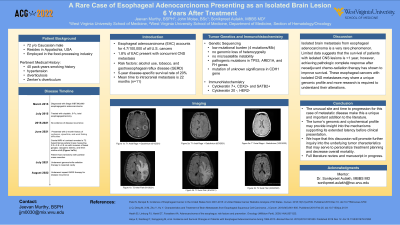
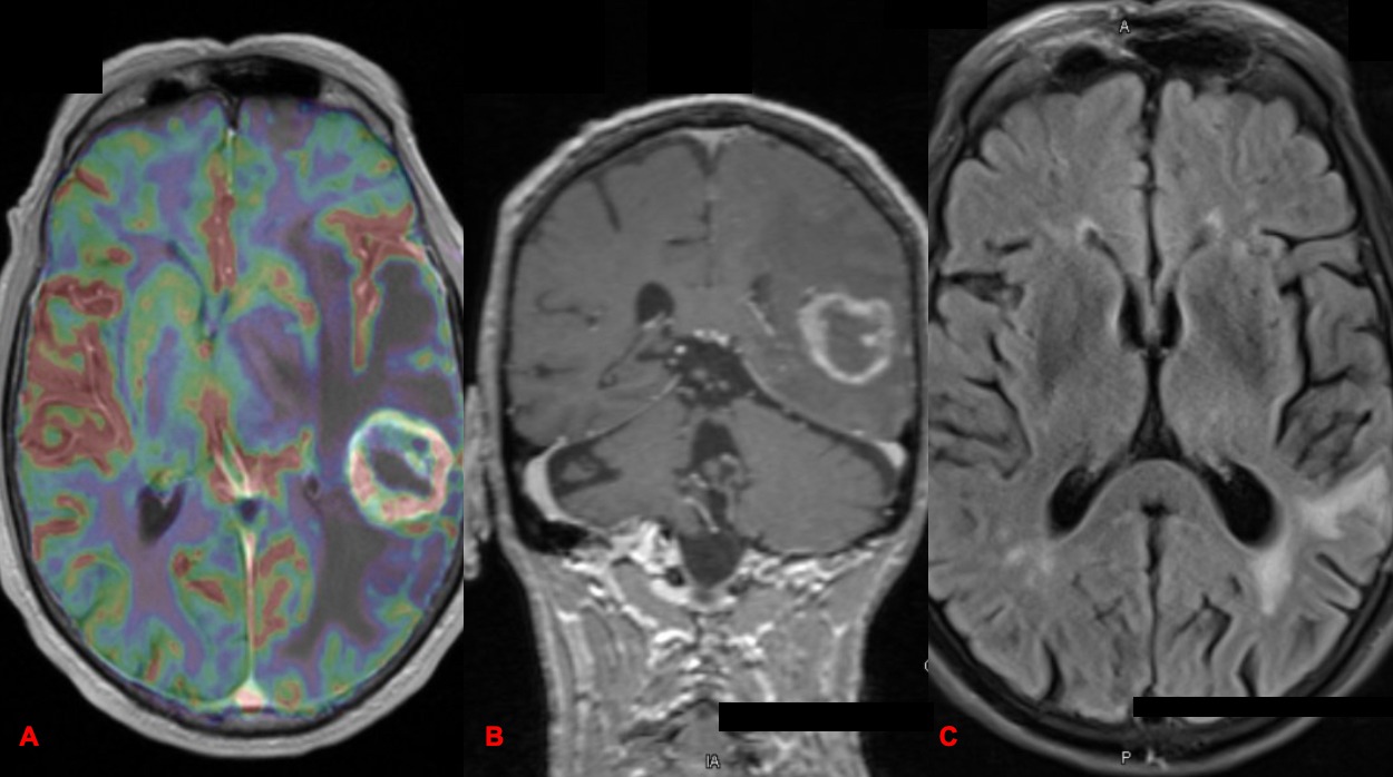
Case Description/Methods: A 72-year-old-male with a 40 pack-year smoking presented with progressive dysphagia in March 2015. Contrast enhanced CT of the chest, abdomen and pelvis revealed a constricting mass at the esophagogastric junction that led to endoscopy guided biopsy revealing esophageal adenocarcinoma. He underwent neoadjuvant chemotherapy (cisplatin/5-fluorouracil) and radiation therapy followed by esophagogastrectomy in July of 2015 where diagnosis of stage IIIBT3N2cM0 esophageal adenocarcinoma was confirmed. Clinical and imaging surveillance for 6 years had not shown any progression of cancer. However, in June of 2021, he presented with altered mental status that led to contrast enhanced brain MRI showing a T2 hyperintense left parietal lobe mass with an 8 mm midline shift and surrounding vasogenic edema. FDG PET-CT head to toes did not show extracranial recurrence. He underwent left posterior temporal craniotomy for resection of tumor confirming the diagnosis of metastatic esophageal adenocarcinoma. IHC showed positivity of cytokeratin7, CDX2 and SATB2. Whole exome sequencing revealed mutations in ARID1A, FH, and TP53 with variants of unknown significance of ATM and CDH. Tumor displayed proficient mismatch repair protein, a combined positive score of 15 for PD-L1, and negative Her2/Neu. Further sequencing showed tumor mutational burden of 4, no genomic loss of heterozygosity, and stable microsatellite repeats. He received gamma-knife radiation to the solitary left temporal lobe lesion. Patient continues to maintain his quality of life with no evidence of clinical and radiographic recurrence.
Discussion: Isolated brain metastasis from esophageal adenocarcinoma is a very rare phenomenon. Limited data suggests that the survival of patients with isolated CNS lesions is < 1 year, however, achieving pathologic complete response after neoadjuvant chemo-radiation therapy has shown to improve survival. These esophageal cancers with isolated CNS metastases may share a unique genomic profile and more research is required to understand their alterations.

Disclosures:
Jeevan J. Murthy, BSPH1, John Moise, BSc1, Sonikpreet Aulakh, MBBS, MD2. E0248 - A Rare Case of Esophageal Adenocarcinoma Presenting as an Isolated Brain Lesion 6 Years After Treatment, ACG 2022 Annual Scientific Meeting Abstracts. Charlotte, NC: American College of Gastroenterology.
1West Virginia University School of Medicine, Morgantown, WV; 2West Virginia University, Morgantown, WV
Introduction: Esophageal adenocarcinomas account for ~1% of all cancers in the U.S. The prevalence of central nervous system (CNS) metastases associated with these cancers is around 1.8%. Response rates to standard of care are poor and prognosis is dismal. Often CNS involvement is associated with widely metastatic disease, herein, we present a case of a solitary brain lesion from esophageal adenocarcinoma at first recurrence.
Case Description/Methods: A 72-year-old-male with a 40 pack-year smoking presented with progressive dysphagia in March 2015. Contrast enhanced CT of the chest, abdomen and pelvis revealed a constricting mass at the esophagogastric junction that led to endoscopy guided biopsy revealing esophageal adenocarcinoma. He underwent neoadjuvant chemotherapy (cisplatin/5-fluorouracil) and radiation therapy followed by esophagogastrectomy in July of 2015 where diagnosis of stage IIIBT3N2cM0 esophageal adenocarcinoma was confirmed. Clinical and imaging surveillance for 6 years had not shown any progression of cancer. However, in June of 2021, he presented with altered mental status that led to contrast enhanced brain MRI showing a T2 hyperintense left parietal lobe mass with an 8 mm midline shift and surrounding vasogenic edema. FDG PET-CT head to toes did not show extracranial recurrence. He underwent left posterior temporal craniotomy for resection of tumor confirming the diagnosis of metastatic esophageal adenocarcinoma. IHC showed positivity of cytokeratin7, CDX2 and SATB2. Whole exome sequencing revealed mutations in ARID1A, FH, and TP53 with variants of unknown significance of ATM and CDH. Tumor displayed proficient mismatch repair protein, a combined positive score of 15 for PD-L1, and negative Her2/Neu. Further sequencing showed tumor mutational burden of 4, no genomic loss of heterozygosity, and stable microsatellite repeats. He received gamma-knife radiation to the solitary left temporal lobe lesion. Patient continues to maintain his quality of life with no evidence of clinical and radiographic recurrence.
Discussion: Isolated brain metastasis from esophageal adenocarcinoma is a very rare phenomenon. Limited data suggests that the survival of patients with isolated CNS lesions is < 1 year, however, achieving pathologic complete response after neoadjuvant chemo-radiation therapy has shown to improve survival. These esophageal cancers with isolated CNS metastases may share a unique genomic profile and more research is required to understand their alterations.

Figure: MRI of the brain, Figure 1A- Pre-op axial OLEA of L. temporal lobe tumor (06/2021), 1B- Pre-op axial coronal T1 (06/2021), 1C- Follow-up T2 MRI
Disclosures:
Jeevan Murthy indicated no relevant financial relationships.
John Moise indicated no relevant financial relationships.
Sonikpreet Aulakh: Novocure – Speakers Bureau. TEMPUS – Advisor or Review Panel Member.
Jeevan J. Murthy, BSPH1, John Moise, BSc1, Sonikpreet Aulakh, MBBS, MD2. E0248 - A Rare Case of Esophageal Adenocarcinoma Presenting as an Isolated Brain Lesion 6 Years After Treatment, ACG 2022 Annual Scientific Meeting Abstracts. Charlotte, NC: American College of Gastroenterology.

