Back


Poster Session E - Tuesday Afternoon
Category: General Endoscopy
E0287 - Limitations of Endoscopic Tools for Foreign Body Retrieval: A Case of Massive Ferromagnet Ingestion
Tuesday, October 25, 2022
3:00 PM – 5:00 PM ET
Location: Crown Ballroom

Has Audio
- PS
Pir Shah, MD
University of Texas Health Science Center
San Antonio, Texas
Presenting Author(s)
Pir Ahmad Shah, MD1, Bara El Kurdi, MD2, Jason Rocha, MD2
1University of Texas Health Science Center, San Antonio, TX; 2UTHSCSA, San Antonio, TX
Introduction: Esophagogastroduodenoscopy (EGD) remains the standard of care for foreign body (FB) retrieval. The decision to pursue endoscopic intervention is based on multiple considerations including the probability of passage of the FB, the risk of potential injury caused by ingestion of the foreign body, and the risk of potential injury caused by performing the intervention. We present a case of FB ingestion resulting in practically impossible endoscopic retrieval. We also highlight endoscopic cues that should prompt a gastroenterologist to consider surgical evaluation in high-risk cases.
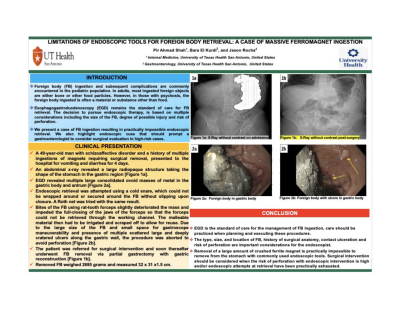
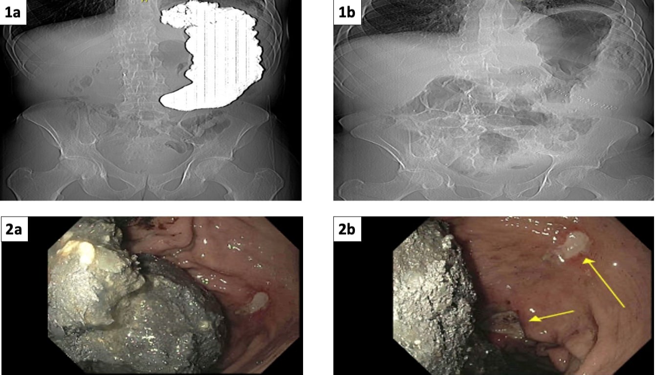
Case Description/Methods: A 49-year-old man with schizoaffective disorder and a history of multiple ingestions of foreign bodies presented to the hospital for vomiting and diarrhea for 4 days. An abdominal x-ray revealed a large radiopaque structure that conformed to the shape of the stomach in the gastric region [Figure 1a]. EGD revealed multiple large consolidated ovoid masses of gray metal in the gastric body and antrum [Figure 2a]. Endoscopic retrieval was attempted using a large cold snare, which could not be wrapped around or secured around the FB without slipping upon closure. A Roth net was tried with the same unsuccessful result. Bites of the FB using rat-tooth forceps slightly deteriorated the mass and the foreign body material impeded the full-closing of the jaws of the forceps so that the forceps could not be retrieved through the working channel. The malleable material then had to be irrigated and scraped off to allow for reuse. Due to the large size of the FB and small space in the stomach for gastroscope maneuverability and the presence of multiple scattered large and deeply cratered ulcers along the gastric wall, the procedure was aborted to avoid perforation [Figure 2b]. The patient was referred for surgical intervention and soon thereafter underwent FB removal via partial gastrectomy with gastric reconstruction [Figure 1b]. The recovered FB weighed 2,885 grams and measured 32 x 31 x 1.5 cm
Discussion: Care should be practiced when planning and executing the retrieval of foreign bodies. The type, size, and location of the FB, the patient’s anatomy, the presence of deep ulceration and the risk of organ perforation are important considerations for the endoscopist. Surgical intervention should be considered when the risk of perforation with endoscopic intervention is high and/or endoscopic attempts at retrieval have been unsuccessful after practically exhausting the endoscopic tools available.

Disclosures:
Pir Ahmad Shah, MD1, Bara El Kurdi, MD2, Jason Rocha, MD2. E0287 - Limitations of Endoscopic Tools for Foreign Body Retrieval: A Case of Massive Ferromagnet Ingestion, ACG 2022 Annual Scientific Meeting Abstracts. Charlotte, NC: American College of Gastroenterology.
1University of Texas Health Science Center, San Antonio, TX; 2UTHSCSA, San Antonio, TX
Introduction: Esophagogastroduodenoscopy (EGD) remains the standard of care for foreign body (FB) retrieval. The decision to pursue endoscopic intervention is based on multiple considerations including the probability of passage of the FB, the risk of potential injury caused by ingestion of the foreign body, and the risk of potential injury caused by performing the intervention. We present a case of FB ingestion resulting in practically impossible endoscopic retrieval. We also highlight endoscopic cues that should prompt a gastroenterologist to consider surgical evaluation in high-risk cases.
Case Description/Methods: A 49-year-old man with schizoaffective disorder and a history of multiple ingestions of foreign bodies presented to the hospital for vomiting and diarrhea for 4 days. An abdominal x-ray revealed a large radiopaque structure that conformed to the shape of the stomach in the gastric region [Figure 1a]. EGD revealed multiple large consolidated ovoid masses of gray metal in the gastric body and antrum [Figure 2a]. Endoscopic retrieval was attempted using a large cold snare, which could not be wrapped around or secured around the FB without slipping upon closure. A Roth net was tried with the same unsuccessful result. Bites of the FB using rat-tooth forceps slightly deteriorated the mass and the foreign body material impeded the full-closing of the jaws of the forceps so that the forceps could not be retrieved through the working channel. The malleable material then had to be irrigated and scraped off to allow for reuse. Due to the large size of the FB and small space in the stomach for gastroscope maneuverability and the presence of multiple scattered large and deeply cratered ulcers along the gastric wall, the procedure was aborted to avoid perforation [Figure 2b]. The patient was referred for surgical intervention and soon thereafter underwent FB removal via partial gastrectomy with gastric reconstruction [Figure 1b]. The recovered FB weighed 2,885 grams and measured 32 x 31 x 1.5 cm
Discussion: Care should be practiced when planning and executing the retrieval of foreign bodies. The type, size, and location of the FB, the patient’s anatomy, the presence of deep ulceration and the risk of organ perforation are important considerations for the endoscopist. Surgical intervention should be considered when the risk of perforation with endoscopic intervention is high and/or endoscopic attempts at retrieval have been unsuccessful after practically exhausting the endoscopic tools available.

Figure: Figure 1a: CT scan without contrast on admission
Figure 1b: CT scan without contrast post-surgery
Figure 2a: Foreign body in gastric body
Figure 2b: Foreign body with ulcers in gastric body
Figure 1b: CT scan without contrast post-surgery
Figure 2a: Foreign body in gastric body
Figure 2b: Foreign body with ulcers in gastric body
Disclosures:
Pir Ahmad Shah indicated no relevant financial relationships.
Bara El Kurdi indicated no relevant financial relationships.
Jason Rocha indicated no relevant financial relationships.
Pir Ahmad Shah, MD1, Bara El Kurdi, MD2, Jason Rocha, MD2. E0287 - Limitations of Endoscopic Tools for Foreign Body Retrieval: A Case of Massive Ferromagnet Ingestion, ACG 2022 Annual Scientific Meeting Abstracts. Charlotte, NC: American College of Gastroenterology.
