Back


Poster Session E - Tuesday Afternoon
Category: GI Bleeding
E0323 - Retroperitoneal Leiomyosarcoma: An Unusual Cause of Gastrointestinal Bleeding
Tuesday, October 25, 2022
3:00 PM – 5:00 PM ET
Location: Crown Ballroom

Has Audio
.jpg)
Baruh B. Mulat
Montefiore Medical Center
Bronx, NY
Presenting Author(s)
Baruh B. Mulat, , Tehseen Haider, MD, Harish Guddati, MD,
Montefiore Medical Center, Bronx, NY
Introduction: Retroperitoneal leiomyosarcoma (RLMS) is a malignant tumor originating from a smooth muscle tissue, and is rare with an incidence rate of 2 per million population. The large retroperitoneal space allows slow growth of the tumor to significant size before compressive symptoms arise. Diagnosis of such tumors as a result of a tumor invasion to the gastrointestinal tract is rare. We herein present a case of a patient diagnosed with a RLMS invading the duodenum and presenting with gastrointestinal bleeding.
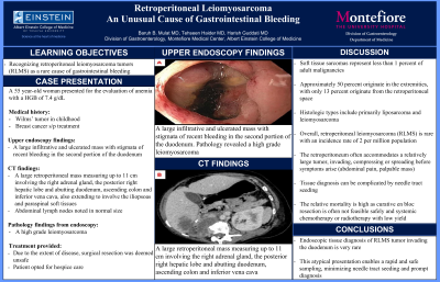
Case Description/Methods: A 55 year-old woman presented to the emergency department for the evaluation of anemia with a HGB of 7.4 g/dL. Past medical history of Willms tumor and breast cancer was noted. Upper endoscopy revealed a large infiltrative and ulcerated mass with stigmata of recent bleeding in the second portion of the duodenum. Colonoscopy was unremarkable. Computed tomography revealed a large retroperitoneal mass measuring up to 11 cm involving the right adrenal gland, the posterior right hepatic lobe and abutting duodenum, ascending colon and inferior vena cava, also extending to involve the iliopsoas and paraspinal soft tissues. Abdominal lymph nodes noted in normal size. Surgical pathology from the duodenal mass revealed a high grade leiomyosarcoma. Due to the extent of disease, surgical resection was deemed unsafe. Unfortunately the patient died before chemotherapy initiation.
Discussion: Soft tissue sarcomas represent less than 1 percent of adult malignancies. Approximately 50 percent originate in the extremities, with only 13 percent originate from the retroperitoneal space. Histologic types include primarily liposarcoma and leiomyosarcoma. Overall, retroperitoneal leiomyosarcoma (RLMS) is rare with an incidence rate of 2 per million population. The retroperitoneum often accommodates a relatively large tumor before symptoms arise. Non specific abdominal pain and palpable mass can be the presentation. Abdominal imaging often reveals a large retroperitoneal tumor, compressing, invading or spreading metastasis to organs or structures. Careful attention is needed to avoid needle tract seeding during tissue sampling. The relative mortality is high as curative en bloc resection is often not feasible safely. Systemic chemotherapy or radiotherapy have been reported of low yield in advanced, unresectable stages. Endoscopic tissue diagnosis from RLMS tumors invading the duodenum is very rare. The atypical presentation described herin enabled a rapid and safe sampling.

Disclosures:
Baruh B. Mulat, , Tehseen Haider, MD, Harish Guddati, MD, . E0323 - Retroperitoneal Leiomyosarcoma: An Unusual Cause of Gastrointestinal Bleeding, ACG 2022 Annual Scientific Meeting Abstracts. Charlotte, NC: American College of Gastroenterology.
Montefiore Medical Center, Bronx, NY
Introduction: Retroperitoneal leiomyosarcoma (RLMS) is a malignant tumor originating from a smooth muscle tissue, and is rare with an incidence rate of 2 per million population. The large retroperitoneal space allows slow growth of the tumor to significant size before compressive symptoms arise. Diagnosis of such tumors as a result of a tumor invasion to the gastrointestinal tract is rare. We herein present a case of a patient diagnosed with a RLMS invading the duodenum and presenting with gastrointestinal bleeding.
Case Description/Methods: A 55 year-old woman presented to the emergency department for the evaluation of anemia with a HGB of 7.4 g/dL. Past medical history of Willms tumor and breast cancer was noted. Upper endoscopy revealed a large infiltrative and ulcerated mass with stigmata of recent bleeding in the second portion of the duodenum. Colonoscopy was unremarkable. Computed tomography revealed a large retroperitoneal mass measuring up to 11 cm involving the right adrenal gland, the posterior right hepatic lobe and abutting duodenum, ascending colon and inferior vena cava, also extending to involve the iliopsoas and paraspinal soft tissues. Abdominal lymph nodes noted in normal size. Surgical pathology from the duodenal mass revealed a high grade leiomyosarcoma. Due to the extent of disease, surgical resection was deemed unsafe. Unfortunately the patient died before chemotherapy initiation.
Discussion: Soft tissue sarcomas represent less than 1 percent of adult malignancies. Approximately 50 percent originate in the extremities, with only 13 percent originate from the retroperitoneal space. Histologic types include primarily liposarcoma and leiomyosarcoma. Overall, retroperitoneal leiomyosarcoma (RLMS) is rare with an incidence rate of 2 per million population. The retroperitoneum often accommodates a relatively large tumor before symptoms arise. Non specific abdominal pain and palpable mass can be the presentation. Abdominal imaging often reveals a large retroperitoneal tumor, compressing, invading or spreading metastasis to organs or structures. Careful attention is needed to avoid needle tract seeding during tissue sampling. The relative mortality is high as curative en bloc resection is often not feasible safely. Systemic chemotherapy or radiotherapy have been reported of low yield in advanced, unresectable stages. Endoscopic tissue diagnosis from RLMS tumors invading the duodenum is very rare. The atypical presentation described herin enabled a rapid and safe sampling.

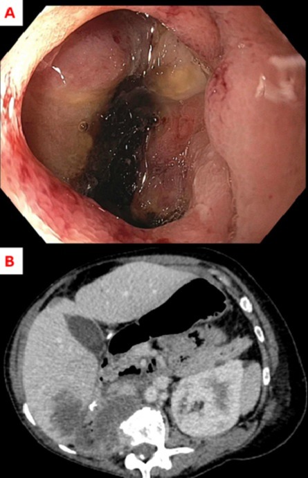
Figure: A - Endoscopic findings. A large infiltrative and ulcerated mass with stigmata of recent bleeding in the second portion of the duodenum. Surgical pathology revealed a high grade leiomyosarcoma.
B - Computed tomography of the abdomen and pelvis. Large retroperitoneal mass measuring up to 11 cm involving the right adrenal gland, the posterior right hepatic lobe and abutting duodenum, ascending colon and inferior vena cava, also extending to involve the iliopsoas and paraspinal soft tissues.
B - Computed tomography of the abdomen and pelvis. Large retroperitoneal mass measuring up to 11 cm involving the right adrenal gland, the posterior right hepatic lobe and abutting duodenum, ascending colon and inferior vena cava, also extending to involve the iliopsoas and paraspinal soft tissues.
Disclosures:
Baruh Mulat indicated no relevant financial relationships.
Tehseen Haider indicated no relevant financial relationships.
Harish Guddati, MD indicated no relevant financial relationships.
Baruh B. Mulat, , Tehseen Haider, MD, Harish Guddati, MD, . E0323 - Retroperitoneal Leiomyosarcoma: An Unusual Cause of Gastrointestinal Bleeding, ACG 2022 Annual Scientific Meeting Abstracts. Charlotte, NC: American College of Gastroenterology.
