Back


Poster Session A - Sunday Afternoon
Category: Esophagus
A0245 - A Case Report of Iatrogenic Esophageal Perforation: Complication of Orogastric Tube Placement
Sunday, October 23, 2022
5:00 PM – 7:00 PM ET
Location: Crown Ballroom

Has Audio

Ashik Pokharel, MBBS
MedStar Health
Baltimore, MD
Presenting Author(s)
Ashik Pokharel, MBBS, Yogesh Subedi, MBBS, Kalyan Paudel, MD
MedStar Health, Baltimore, MD
Introduction: Orogastric tube insertion is a routine procedure in medical care. However, misplacement of the tube can cause a variety of complications, which can be life threatening in some instances.
Case Description/Methods: 71-year-old male presented with dyspnea, fever, chills, cough, and myalgia for 2 weeks. He had tachycardia, tachypnea, and was hypoxic to 66% in room air. He was found to have acute hypoxic respiratory failure secondary to COVID 19 Pneumonia and was admitted to ICU. But, he continued to be hypoxic and was started on BiPAP. He eventually became altered, and was intubated. Post intubation orogastric tube (OGT) placement was unsuccessful on the first attempt due to resistance. On the second attempt, the nurse was able to advance partially. But, a chest XR showed OGT in the mediastinum, and OGT was removed. CT of neck and chest revealed pneumomediastinum with possible mid-thoracic esophageal perforation. The patient was started on broad-spectrum antibiotics and thoracic surgery was consulted. Given his mechanical ventilation requirement, surgery deemed him unfit to tolerate thoracotomy and the endoscopic procedure was not available in the hospital. So, recommendation was to manage conservatively. His hospital course was complicated by hypotension requiring vasopressors and metabolic acidosis in setting of acute renal failure requiring CRRT. Code status was changed by the family to Do Not Resuscitate due to his deteriorating condition. Eventually, he had a PEA arrest and was expired.
Discussion: OGT intubation is performed at hospitals for feeding, medication administration or gastric decompression. Although it is considered a safe procedure, complications can arise due to OGT misplacement or trauma caused by the OGT itself or the intubation process. OGT misplacement is typically endotracheal or intracranial. Misplacement within the upper GI lumen is usually detected by a kink in the oropharynx or esophagus. The subsequent complications are identified by the structure that is perforated (e.g., mediastinitis or pneumothorax). Regardless of whether counteraction is perceived, the physician must be careful not to apply excessive force. The location of the OGT tip should be determined by a chest radiograph; visualization of the tip below the diaphragm verifies appropriate placement. Complications of OGT insertion are uncommon; however, the consequences are potentially serious, and the anatomy of the upper GI tract should be understood by all who are involved in the care.

Disclosures:
Ashik Pokharel, MBBS, Yogesh Subedi, MBBS, Kalyan Paudel, MD. A0245 - A Case Report of Iatrogenic Esophageal Perforation: Complication of Orogastric Tube Placement, ACG 2022 Annual Scientific Meeting Abstracts. Charlotte, NC: American College of Gastroenterology.
MedStar Health, Baltimore, MD
Introduction: Orogastric tube insertion is a routine procedure in medical care. However, misplacement of the tube can cause a variety of complications, which can be life threatening in some instances.
Case Description/Methods: 71-year-old male presented with dyspnea, fever, chills, cough, and myalgia for 2 weeks. He had tachycardia, tachypnea, and was hypoxic to 66% in room air. He was found to have acute hypoxic respiratory failure secondary to COVID 19 Pneumonia and was admitted to ICU. But, he continued to be hypoxic and was started on BiPAP. He eventually became altered, and was intubated. Post intubation orogastric tube (OGT) placement was unsuccessful on the first attempt due to resistance. On the second attempt, the nurse was able to advance partially. But, a chest XR showed OGT in the mediastinum, and OGT was removed. CT of neck and chest revealed pneumomediastinum with possible mid-thoracic esophageal perforation. The patient was started on broad-spectrum antibiotics and thoracic surgery was consulted. Given his mechanical ventilation requirement, surgery deemed him unfit to tolerate thoracotomy and the endoscopic procedure was not available in the hospital. So, recommendation was to manage conservatively. His hospital course was complicated by hypotension requiring vasopressors and metabolic acidosis in setting of acute renal failure requiring CRRT. Code status was changed by the family to Do Not Resuscitate due to his deteriorating condition. Eventually, he had a PEA arrest and was expired.
Discussion: OGT intubation is performed at hospitals for feeding, medication administration or gastric decompression. Although it is considered a safe procedure, complications can arise due to OGT misplacement or trauma caused by the OGT itself or the intubation process. OGT misplacement is typically endotracheal or intracranial. Misplacement within the upper GI lumen is usually detected by a kink in the oropharynx or esophagus. The subsequent complications are identified by the structure that is perforated (e.g., mediastinitis or pneumothorax). Regardless of whether counteraction is perceived, the physician must be careful not to apply excessive force. The location of the OGT tip should be determined by a chest radiograph; visualization of the tip below the diaphragm verifies appropriate placement. Complications of OGT insertion are uncommon; however, the consequences are potentially serious, and the anatomy of the upper GI tract should be understood by all who are involved in the care.

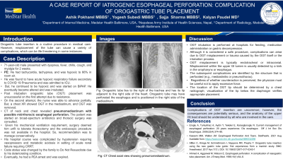
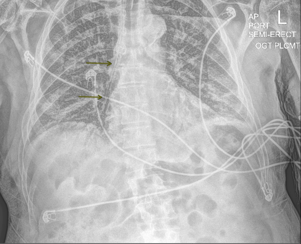
Figure: Orogastric tube lies to the right of the trachea and has its tip adjacent to the right side of the heart. Orogastric tube may have perforated the esophagus and is positioned in the right side of the mediastinum.
Disclosures:
Ashik Pokharel indicated no relevant financial relationships.
Yogesh Subedi indicated no relevant financial relationships.
Kalyan Paudel indicated no relevant financial relationships.
Ashik Pokharel, MBBS, Yogesh Subedi, MBBS, Kalyan Paudel, MD. A0245 - A Case Report of Iatrogenic Esophageal Perforation: Complication of Orogastric Tube Placement, ACG 2022 Annual Scientific Meeting Abstracts. Charlotte, NC: American College of Gastroenterology.
