Back


Poster Session C - Monday Afternoon
Category: Esophagus
C0247 - HSV Esophagitis With Hematemesis and Recalcitrant Food Bolus
Monday, October 24, 2022
3:00 PM – 5:00 PM ET
Location: Crown Ballroom

Has Audio

Tiberiu Moga, MD
University at Buffalo
Buffalo, NY
Presenting Author(s)
Tiberiu Moga, MD1, Kevin Robillard, MD2, Anoop Prabhu, MD2, Sehrish Jamot, MD2, Andrew Bain, MD2
1University at Buffalo, Buffalo, NY; 2Roswell Park Comprehensive Cancer Center, Buffalo, NY
Introduction: HSV esophagitis (HSV-E) affects primarily immunosuppressed individuals. Common symptoms include fever, odynophagia, dysphagia and retrosternal chest pain. Rare manifestations include hematemesis and food bolus. We present a 58 year-old woman with AML refractory to hematopoietic stem cell transplants (HSCTs) who developed HSV-E with hematemesis and food bolus.
Case Description/Methods: A 58 year-old woman presented to an outside hospital with acute dysphagia and hematemesis. Her past medical history includes refractory AML with HSCTs in 2007, 2010 and 2019 and GI graft-versus-host-disease (GVHD) on talazoparib and gemtuzumab. EGD reported possible hematoma in the entire esophagus with superficial mucosal tears. Biopsies were negative for viral cytopathic effect or malignancy. She was transferred to our hospital for NK cell infusion for AML. She had a prolonged hospital course with neutropenic fever and sepsis and was on prophylactic acyclovir.
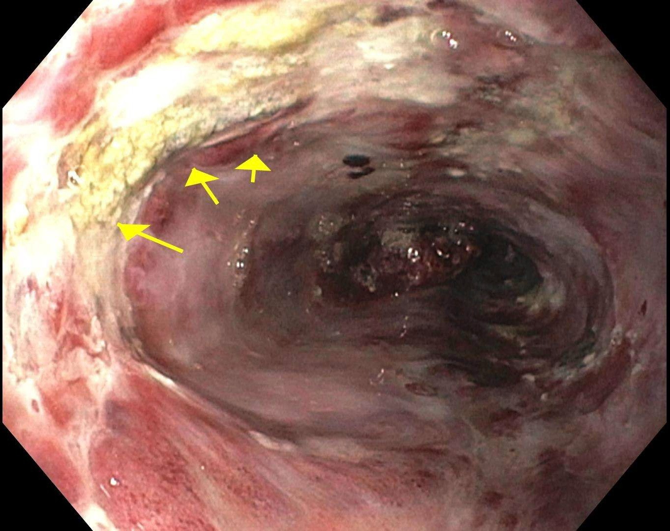
She had dysphagia and EGD week 1 of her admission showed ulcerated, hemorrhagic esophageal mucosa with positive HSV biopsies. She was given IV foscarnet, acyclovir, pantoprazole and sucralfate. EGD was repeated week 3 for persistent dysphagia and odynophagia. Her esophagitis improved and biopsies were again HSV-positive. She had recurrent dysphagia and hematemesis at week 5. An updated EGD showed a massive esophageal food bolus that could not be cleared despite a 4 hour procedure. She was given a trial of Coca-Cola mixed with Creon to help dissolve the bolus. A 4th EGD was done 4 days later. This cleared the bolus after 4 more hours of procedure time.
EGD was repeated at week 13 for foreign body sensation and showed mild sloughing of mucosa at 22 cm but the mucosa was otherwise healed. Biopsies showed squamous esophageal mucosa with chronic inflammation (HSV-negative). She developed recurrent colonic GVHD treated with immunosuppressants. She continued HSV prophylaxis with valganciclovir and did not develop recurrent HSV-E despite ongoing immunosuppression.
Discussion: HSV-E was first described in 1943 and only 1 case of HSV esophagitis presenting with food bolus has been reported. HSV-E most commonly affects immunosuppressed patients, including HSCT patients. The highest risk for HSV reactivation appears to be within 30 days of HSCT (67% of reinfections). Age >50 is associated with reactivation higher risk and HSV reactivation in the setting of cancer is associated with decreased overall survival.

Disclosures:
Tiberiu Moga, MD1, Kevin Robillard, MD2, Anoop Prabhu, MD2, Sehrish Jamot, MD2, Andrew Bain, MD2. C0247 - HSV Esophagitis With Hematemesis and Recalcitrant Food Bolus, ACG 2022 Annual Scientific Meeting Abstracts. Charlotte, NC: American College of Gastroenterology.
1University at Buffalo, Buffalo, NY; 2Roswell Park Comprehensive Cancer Center, Buffalo, NY
Introduction: HSV esophagitis (HSV-E) affects primarily immunosuppressed individuals. Common symptoms include fever, odynophagia, dysphagia and retrosternal chest pain. Rare manifestations include hematemesis and food bolus. We present a 58 year-old woman with AML refractory to hematopoietic stem cell transplants (HSCTs) who developed HSV-E with hematemesis and food bolus.
Case Description/Methods: A 58 year-old woman presented to an outside hospital with acute dysphagia and hematemesis. Her past medical history includes refractory AML with HSCTs in 2007, 2010 and 2019 and GI graft-versus-host-disease (GVHD) on talazoparib and gemtuzumab. EGD reported possible hematoma in the entire esophagus with superficial mucosal tears. Biopsies were negative for viral cytopathic effect or malignancy. She was transferred to our hospital for NK cell infusion for AML. She had a prolonged hospital course with neutropenic fever and sepsis and was on prophylactic acyclovir.
She had dysphagia and EGD week 1 of her admission showed ulcerated, hemorrhagic esophageal mucosa with positive HSV biopsies. She was given IV foscarnet, acyclovir, pantoprazole and sucralfate. EGD was repeated week 3 for persistent dysphagia and odynophagia. Her esophagitis improved and biopsies were again HSV-positive. She had recurrent dysphagia and hematemesis at week 5. An updated EGD showed a massive esophageal food bolus that could not be cleared despite a 4 hour procedure. She was given a trial of Coca-Cola mixed with Creon to help dissolve the bolus. A 4th EGD was done 4 days later. This cleared the bolus after 4 more hours of procedure time.
EGD was repeated at week 13 for foreign body sensation and showed mild sloughing of mucosa at 22 cm but the mucosa was otherwise healed. Biopsies showed squamous esophageal mucosa with chronic inflammation (HSV-negative). She developed recurrent colonic GVHD treated with immunosuppressants. She continued HSV prophylaxis with valganciclovir and did not develop recurrent HSV-E despite ongoing immunosuppression.
Discussion: HSV-E was first described in 1943 and only 1 case of HSV esophagitis presenting with food bolus has been reported. HSV-E most commonly affects immunosuppressed patients, including HSCT patients. The highest risk for HSV reactivation appears to be within 30 days of HSCT (67% of reinfections). Age >50 is associated with reactivation higher risk and HSV reactivation in the setting of cancer is associated with decreased overall survival.

Figure: Figure 1: Upper GI endoscopy image from week 1 showing hemorrhagic and ulcerated (arrows) esophageal mucosa.
Disclosures:
Tiberiu Moga indicated no relevant financial relationships.
Kevin Robillard indicated no relevant financial relationships.
Anoop Prabhu indicated no relevant financial relationships.
Sehrish Jamot indicated no relevant financial relationships.
Andrew Bain indicated no relevant financial relationships.
Tiberiu Moga, MD1, Kevin Robillard, MD2, Anoop Prabhu, MD2, Sehrish Jamot, MD2, Andrew Bain, MD2. C0247 - HSV Esophagitis With Hematemesis and Recalcitrant Food Bolus, ACG 2022 Annual Scientific Meeting Abstracts. Charlotte, NC: American College of Gastroenterology.

