Back


Poster Session E - Tuesday Afternoon
Category: Interventional Endoscopy
E0445 - Endoscopic Evaluation of Gastric Glomus Tumors
Tuesday, October 25, 2022
3:00 PM – 5:00 PM ET
Location: Crown Ballroom

Has Audio

Alexander Malik, MD
Summa Health System
Akron, OH
Presenting Author(s)
Alexander Malik, MD1, Muhammad Nadeem Yousaf, MD2, Sami Samiullah, MD1, Tahan Veysel, MD2, Yuchu Sun, MD1, Amin Mahdi, MD1, Omer Basar, MD2
1Summa Health System, Akron, OH; 2University of Missouri, Columbia, MO
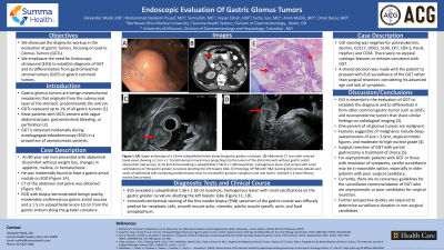
Introduction: Gastric glomus tumors (GGT) are benign mesenchymal neoplasms that originate from the submucosal layer of the stomach, predominantly the antrum, and represent up to 1% of all gastric tumors. Most patients present with vague abdominal pain, gastrointestinal bleeding, or perforation. However, GGT is detected incidentally during esophagogastroduodenoscopy (EGD) in a proportion of asymptomatic patients. Endoscopic ultrasound (EUS) evaluation of GGT is essential to establish the diagnosis and to differentiate it from gastrointestinal stromal tumors (GIST) or gastric carcinoids.
Case Description/Methods: An 80-year-old man presented for abdominal discomfort without weight loss, changes in appetite, melena, or hematochezia. He was incidentally found to have a gastric antral nodule on EGD. EGD with biopsy demonstrated benign patchy, moderately erythematous gastric antral mucosa and a 1.5cm subepithelial lesion 10cm from the gastric antrum along the greater curvature. EUS revealed a subepithelial 1.56cm x 1.28cm isoechoic, homogenous lesion with small calcifications on the gastric greater curvature abutting the left hepatic lobe. Immunohistochemical staining of the fine needle biopsy (FNB) specimen of the gastric nodule was diffusely positive for neoplastic cells, smooth muscle actin, vimentin, patchy muscle specific actin, and focal synaptophysin. Cell staining was negative for pancytokeratin, desmin, CD117, DOG1, S100, CK7, CDX-2, Pax-8, HepPar1 and CD34. There were no atypical cytologic features or mitoses. These findings were consistent with GGT. The patient was not deemed to be a candidate for surgical resection due to advanced age and resolution of his symptoms. A shared decision was made with the patient to proceed with regular surveillance of the GGT rather than surgical resection. He was discharged with a plan of repeat EUS for surveillance of the GGT in one year.
Discussion: The present case illustrated the importance of EUS evaluation of GGT to establish the diagnosis and to differentiate it from other common gastric tumor types. Currently, there are no guideline recommendations for the surveillance of GGT detected on routine EGD in asymptomatic individuals. A definitive surgical treatment with partial gastrectomy was favored in previously published literature. However, for asymptomatic patients with GGT or those with resolution of symptoms, careful surveillance may be a reasonable option, especially in older patients with poor surgical candidacy.

Disclosures:
Alexander Malik, MD1, Muhammad Nadeem Yousaf, MD2, Sami Samiullah, MD1, Tahan Veysel, MD2, Yuchu Sun, MD1, Amin Mahdi, MD1, Omer Basar, MD2. E0445 - Endoscopic Evaluation of Gastric Glomus Tumors, ACG 2022 Annual Scientific Meeting Abstracts. Charlotte, NC: American College of Gastroenterology.
1Summa Health System, Akron, OH; 2University of Missouri, Columbia, MO
Introduction: Gastric glomus tumors (GGT) are benign mesenchymal neoplasms that originate from the submucosal layer of the stomach, predominantly the antrum, and represent up to 1% of all gastric tumors. Most patients present with vague abdominal pain, gastrointestinal bleeding, or perforation. However, GGT is detected incidentally during esophagogastroduodenoscopy (EGD) in a proportion of asymptomatic patients. Endoscopic ultrasound (EUS) evaluation of GGT is essential to establish the diagnosis and to differentiate it from gastrointestinal stromal tumors (GIST) or gastric carcinoids.
Case Description/Methods: An 80-year-old man presented for abdominal discomfort without weight loss, changes in appetite, melena, or hematochezia. He was incidentally found to have a gastric antral nodule on EGD. EGD with biopsy demonstrated benign patchy, moderately erythematous gastric antral mucosa and a 1.5cm subepithelial lesion 10cm from the gastric antrum along the greater curvature. EUS revealed a subepithelial 1.56cm x 1.28cm isoechoic, homogenous lesion with small calcifications on the gastric greater curvature abutting the left hepatic lobe. Immunohistochemical staining of the fine needle biopsy (FNB) specimen of the gastric nodule was diffusely positive for neoplastic cells, smooth muscle actin, vimentin, patchy muscle specific actin, and focal synaptophysin. Cell staining was negative for pancytokeratin, desmin, CD117, DOG1, S100, CK7, CDX-2, Pax-8, HepPar1 and CD34. There were no atypical cytologic features or mitoses. These findings were consistent with GGT. The patient was not deemed to be a candidate for surgical resection due to advanced age and resolution of his symptoms. A shared decision was made with the patient to proceed with regular surveillance of the GGT rather than surgical resection. He was discharged with a plan of repeat EUS for surveillance of the GGT in one year.
Discussion: The present case illustrated the importance of EUS evaluation of GGT to establish the diagnosis and to differentiate it from other common gastric tumor types. Currently, there are no guideline recommendations for the surveillance of GGT detected on routine EGD in asymptomatic individuals. A definitive surgical treatment with partial gastrectomy was favored in previously published literature. However, for asymptomatic patients with GGT or those with resolution of symptoms, careful surveillance may be a reasonable option, especially in older patients with poor surgical candidacy.

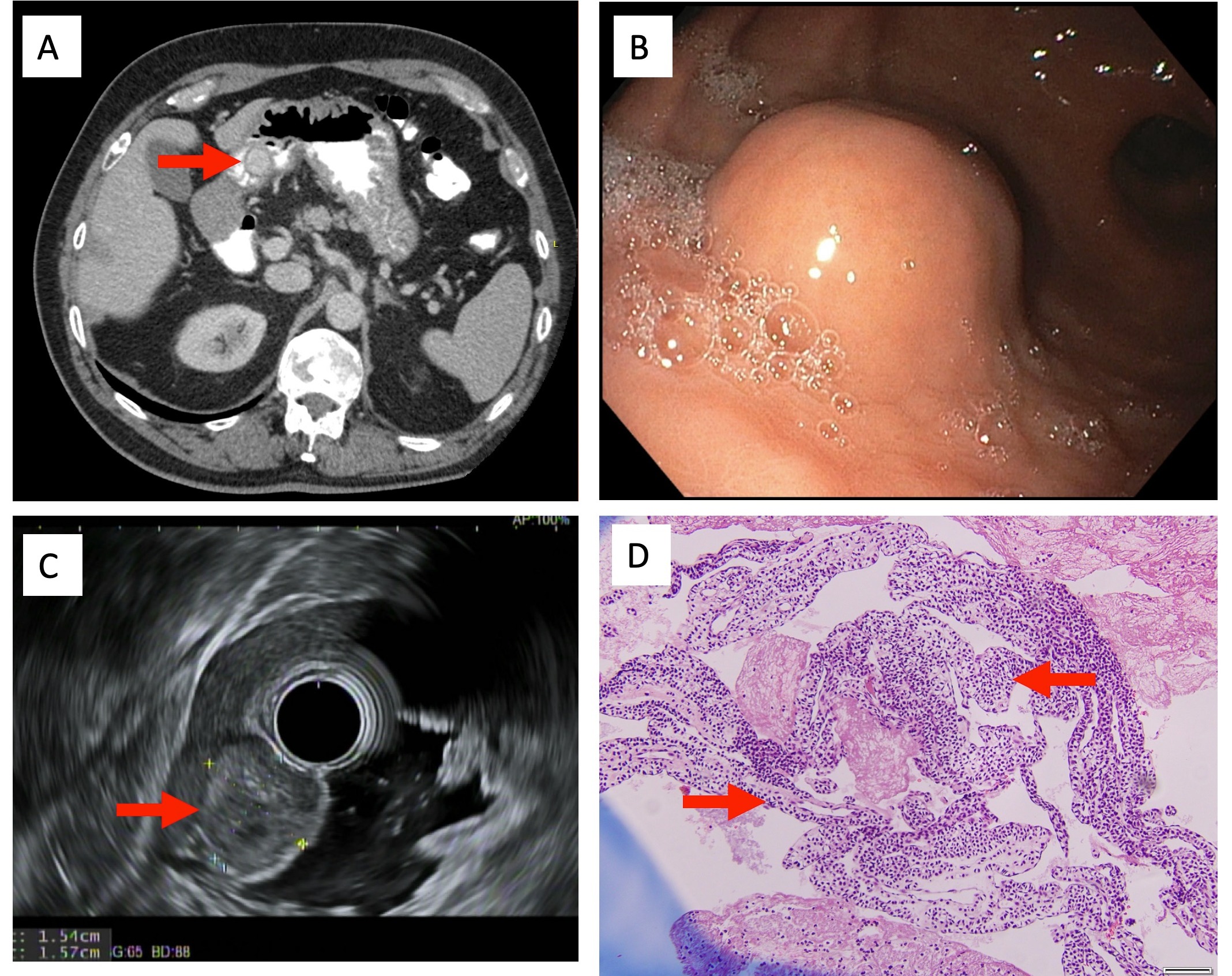
Figure: Figure 1
A. Abdominal CT scan with contrast (axial view) showing a 2.1cm x 1.7cm intraluminal mural mass projecting into the lumen of the distal stomach without gastric outlet obstruction (red arrow).
B. Upper endoscopy of a 15mm subepithelial lesion along the gastric greater curvature.
C. EUS demonstrating a subepithelial 1.56cm x 1.28cm isoechoic, homogenous lesion (red arrow) with small calcifications on the gastric greater curvature abutting the left hepatic lobe.
D. Histology of FNB with H&E staining (10x) shows lobules and cords of epithelioid cells containing predominantly clear to eosinophilic granular cytoplasm and oval nuclei, invested in a loose fibrous stroma (red arrows). No mitotic figures, atypia or necrosis are identified.
Disclosures:
Alexander Malik indicated no relevant financial relationships.
Muhammad Nadeem Yousaf indicated no relevant financial relationships.
Sami Samiullah indicated no relevant financial relationships.
Tahan Veysel indicated no relevant financial relationships.
Tahan Veysel — NO DISCLOSURE DATA.
Yuchu Sun indicated no relevant financial relationships.
Amin Mahdi indicated no relevant financial relationships.
Omer Basar indicated no relevant financial relationships.
Alexander Malik, MD1, Muhammad Nadeem Yousaf, MD2, Sami Samiullah, MD1, Tahan Veysel, MD2, Yuchu Sun, MD1, Amin Mahdi, MD1, Omer Basar, MD2. E0445 - Endoscopic Evaluation of Gastric Glomus Tumors, ACG 2022 Annual Scientific Meeting Abstracts. Charlotte, NC: American College of Gastroenterology.
