Back


Poster Session C - Monday Afternoon
Category: Interventional Endoscopy
C0473 - Gastrohepatic Ligament Bronchogenic Cyst Diagnosed by EUS FNA
Monday, October 24, 2022
3:00 PM – 5:00 PM ET
Location: Crown Ballroom

Has Audio

Ellen C. Tan, DO
OhioHealth Riverside Methodist Hospital
Columbus, OH
Presenting Author(s)
Ellen C. Tan, DO1, David Y. Lo, MD, FACG2
1OhioHealth Riverside Methodist Hospital, Columbus, OH; 2Ohio Gastroenterology Group, Inc, and The Ohio State University College of Medicine, Columbus, OH
Introduction: Bronchogenic cysts pose diagnostic and therapeutic challenges due to its varied locations and presentations.
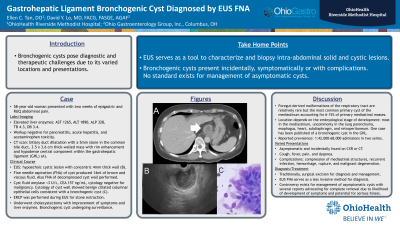
Case Description/Methods: 58-year-old woman presented with two weeks of epigastric and RUQ abdominal pain. Presenting labs: TB 4.3 mg/dL, DB 3.4 mg/dL, AP 328 U/L, AST 1,265 U/L, ALT 1,890 U/L. Workup was negative for pancreatitis, acute hepatitis, and acetaminophen toxicity. CT scan showed biliary duct dilatation with a 5mm stone in the common bile duct, and a 3.5 x 3.6 cm thick-walled mass with rim enhancement and hypodense central component within the gastrohepatic ligament (GHL) (A). EUS characterized the mass as a hypoechoic cystic lesion with a concentric 4mm thick wall (B). Fine needle aspiration (FNA) of the cyst produced 14ml of brown and viscous fluid. After cyst aspirate, FNA of the decompressed cyst wall was also performed. Cyst fluid amylase was < 3 U/L, CEA was 157 ng/mL, and cytology was negative for malignancy. Cytology of the cyst wall showed benign ciliated columnar epithelial cells consistent with a bronchogenic cyst (C). ERCP was performed during EUS for stone extraction. She then underwent cholecystectomy with improvement of symptoms and liver enzymes. Her asymptomatic bronchogenic cyst is undergoing active surveillance.
Discussion: Bronchogenic cysts are foregut-derived malformations of the respiratory tract and while relatively rare, is the most common primary cyst of the mediastinum accounting for 6-15% of primary mediastinal masses. The location depends on the embryological stage of development when the anomaly occurs. Most are in the mediastinum and uncommonly in the lung parenchyma, esophagus, heart, subdiaphragm, and retroperitoneum. Only one other case has been reported of a bronchogenic cyst in the GHL. Reported prevalence is 1:42,000-68,000 admissions in two hospital series. Some present with symptoms such as cough, fever, pain, and dyspnea. Complications can arise with compression of mediastinal structures, recurrent infection, hemorrhage, rupture, and malignant degeneration. Others are asymptomatic and incidentally found on chest radiograph or cross-sectional imaging. No standard exists for treatment. Traditionally, patients underwent surgical excision for definitive diagnosis and management. EUS FNA serves as a less invasive method for diagnosis. Controversy still exists regarding management of asymptomatic cysts with several reports advocating for complete removal due to likelihood of development of symptoms and potential for serious illness.

Disclosures:
Ellen C. Tan, DO1, David Y. Lo, MD, FACG2. C0473 - Gastrohepatic Ligament Bronchogenic Cyst Diagnosed by EUS FNA, ACG 2022 Annual Scientific Meeting Abstracts. Charlotte, NC: American College of Gastroenterology.
1OhioHealth Riverside Methodist Hospital, Columbus, OH; 2Ohio Gastroenterology Group, Inc, and The Ohio State University College of Medicine, Columbus, OH
Introduction: Bronchogenic cysts pose diagnostic and therapeutic challenges due to its varied locations and presentations.
Case Description/Methods: 58-year-old woman presented with two weeks of epigastric and RUQ abdominal pain. Presenting labs: TB 4.3 mg/dL, DB 3.4 mg/dL, AP 328 U/L, AST 1,265 U/L, ALT 1,890 U/L. Workup was negative for pancreatitis, acute hepatitis, and acetaminophen toxicity. CT scan showed biliary duct dilatation with a 5mm stone in the common bile duct, and a 3.5 x 3.6 cm thick-walled mass with rim enhancement and hypodense central component within the gastrohepatic ligament (GHL) (A). EUS characterized the mass as a hypoechoic cystic lesion with a concentric 4mm thick wall (B). Fine needle aspiration (FNA) of the cyst produced 14ml of brown and viscous fluid. After cyst aspirate, FNA of the decompressed cyst wall was also performed. Cyst fluid amylase was < 3 U/L, CEA was 157 ng/mL, and cytology was negative for malignancy. Cytology of the cyst wall showed benign ciliated columnar epithelial cells consistent with a bronchogenic cyst (C). ERCP was performed during EUS for stone extraction. She then underwent cholecystectomy with improvement of symptoms and liver enzymes. Her asymptomatic bronchogenic cyst is undergoing active surveillance.
Discussion: Bronchogenic cysts are foregut-derived malformations of the respiratory tract and while relatively rare, is the most common primary cyst of the mediastinum accounting for 6-15% of primary mediastinal masses. The location depends on the embryological stage of development when the anomaly occurs. Most are in the mediastinum and uncommonly in the lung parenchyma, esophagus, heart, subdiaphragm, and retroperitoneum. Only one other case has been reported of a bronchogenic cyst in the GHL. Reported prevalence is 1:42,000-68,000 admissions in two hospital series. Some present with symptoms such as cough, fever, pain, and dyspnea. Complications can arise with compression of mediastinal structures, recurrent infection, hemorrhage, rupture, and malignant degeneration. Others are asymptomatic and incidentally found on chest radiograph or cross-sectional imaging. No standard exists for treatment. Traditionally, patients underwent surgical excision for definitive diagnosis and management. EUS FNA serves as a less invasive method for diagnosis. Controversy still exists regarding management of asymptomatic cysts with several reports advocating for complete removal due to likelihood of development of symptoms and potential for serious illness.

Figure: CT, EUS, Cytology
Disclosures:
Ellen Tan indicated no relevant financial relationships.
David Lo indicated no relevant financial relationships.
Ellen C. Tan, DO1, David Y. Lo, MD, FACG2. C0473 - Gastrohepatic Ligament Bronchogenic Cyst Diagnosed by EUS FNA, ACG 2022 Annual Scientific Meeting Abstracts. Charlotte, NC: American College of Gastroenterology.
