Back


Poster Session B - Monday Morning
Category: Biliary/Pancreas
B0082 - Pancreatic Somatostatinoma Associated With Neurofibromatosis Type 1
Monday, October 24, 2022
10:00 AM – 12:00 PM ET
Location: Crown Ballroom

Has Audio

Parth M. Patel, MD
Henry Ford Jackson
Jackson, MI
Presenting Author(s)
Parth M. Patel, MD, Sruthi Ramanan, MD, Harjinder Singh, MD, Priya Menon, MD, Hassan Zreik, MD, Devin Malik, MD
Henry Ford Jackson, Jackson, MI
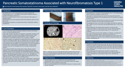
Introduction: Somatostatinoma is a rare neuroendocrine tumor often associated with neurofibromatosis type 1, Von Hippel Lindau syndrome, and tuberous sclerosis. The incidence of somatostatinoma is estimated to be 1 in 40 million. Somatostatinomas have an insidious growth, and as a result, it presents in later stages as a malignant disease. Somatostatinoma presenting with “somatostatinoma syndrome,” which consists of diarrhea, diabetes, and gallstones, is uncommon.
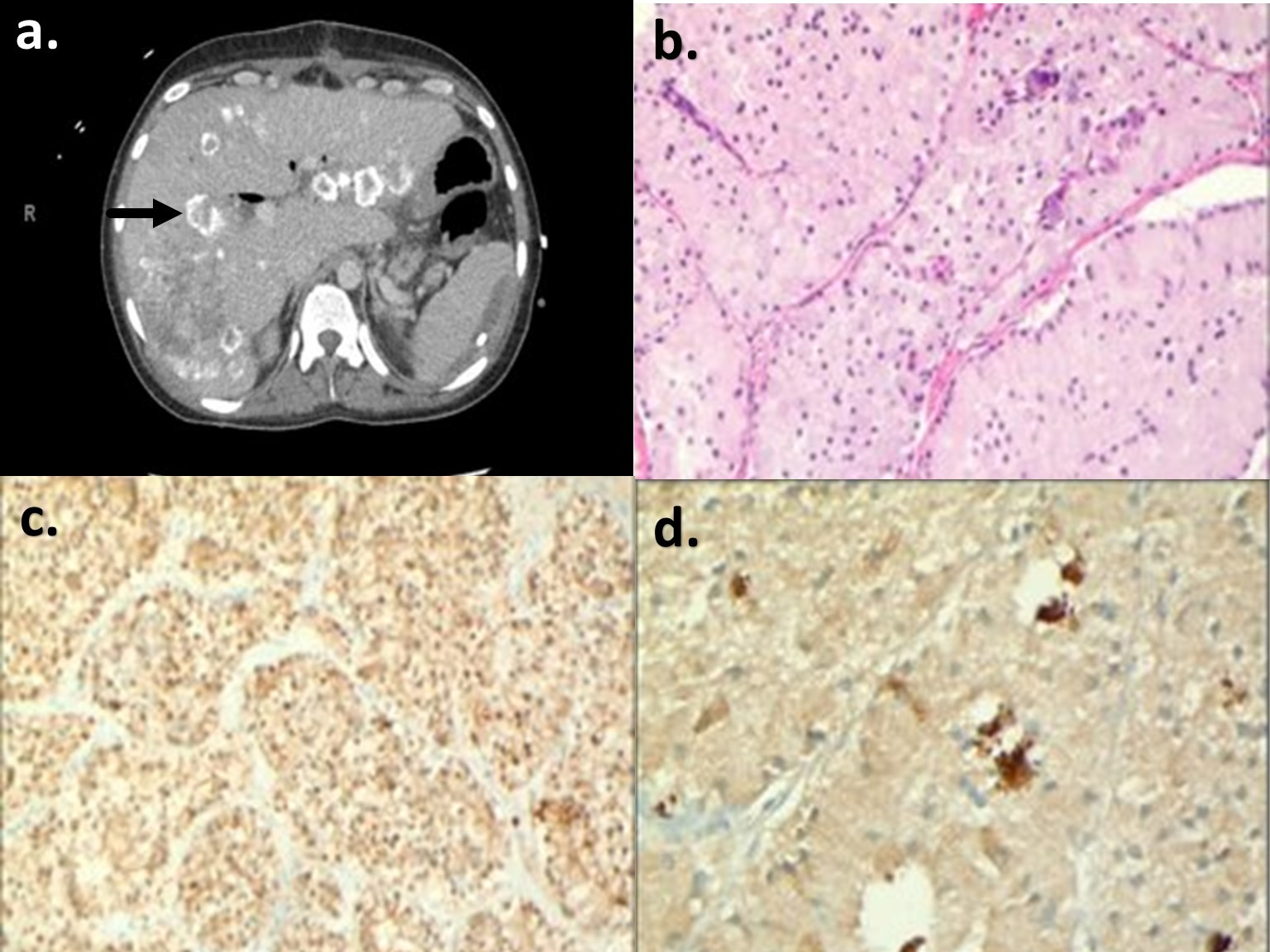
Case Description/Methods: We present the case of a 24-year-old female who presents with newly diagnosed diabetes, diarrhea, and abdominal pain. The patient’s family history consists of MEN (type unknown) syndrome in the patient’s father. Physical examination revealed extensive skin nodules (neurofibromas) and hyperpigmented patches (Café au lait spots), raising concerns for neurofibromatosis. Imaging demonstrated innumerable heterogeneous enhancing masses throughout the hepatic parenchyma and a hypodense lesion in the uncinate process of the pancreas, a stent in the biliary duct with a dilated pancreatic duct, enlarged retroperitoneal and mesenteric lymph nodes, and intraabdominal free-air suggestive of bowel perforation. Biopsy of the liver mass demonstrated neoplastic cells. The immunohistochemical evaluation showed cells that stained positive for AE1/AE3, KI67, Chromogranin, CD56, and CK7. The clinical presentation, radiological appearance, and immunohistochemical staining pattern support a diagnosis of somatostatinoma.
Discussion: Although somatostatinoma presents at a later age, given the insidious growth, it should not be missed in younger individuals with typical symptoms of somatostatinoma syndrome. Given its association with genetic disorders, an effort should be made to distinguish sporadic tumors from those associated with genetic conditions.

Disclosures:
Parth M. Patel, MD, Sruthi Ramanan, MD, Harjinder Singh, MD, Priya Menon, MD, Hassan Zreik, MD, Devin Malik, MD. B0082 - Pancreatic Somatostatinoma Associated With Neurofibromatosis Type 1, ACG 2022 Annual Scientific Meeting Abstracts. Charlotte, NC: American College of Gastroenterology.
Henry Ford Jackson, Jackson, MI
Introduction: Somatostatinoma is a rare neuroendocrine tumor often associated with neurofibromatosis type 1, Von Hippel Lindau syndrome, and tuberous sclerosis. The incidence of somatostatinoma is estimated to be 1 in 40 million. Somatostatinomas have an insidious growth, and as a result, it presents in later stages as a malignant disease. Somatostatinoma presenting with “somatostatinoma syndrome,” which consists of diarrhea, diabetes, and gallstones, is uncommon.
Case Description/Methods: We present the case of a 24-year-old female who presents with newly diagnosed diabetes, diarrhea, and abdominal pain. The patient’s family history consists of MEN (type unknown) syndrome in the patient’s father. Physical examination revealed extensive skin nodules (neurofibromas) and hyperpigmented patches (Café au lait spots), raising concerns for neurofibromatosis. Imaging demonstrated innumerable heterogeneous enhancing masses throughout the hepatic parenchyma and a hypodense lesion in the uncinate process of the pancreas, a stent in the biliary duct with a dilated pancreatic duct, enlarged retroperitoneal and mesenteric lymph nodes, and intraabdominal free-air suggestive of bowel perforation. Biopsy of the liver mass demonstrated neoplastic cells. The immunohistochemical evaluation showed cells that stained positive for AE1/AE3, KI67, Chromogranin, CD56, and CK7. The clinical presentation, radiological appearance, and immunohistochemical staining pattern support a diagnosis of somatostatinoma.
Discussion: Although somatostatinoma presents at a later age, given the insidious growth, it should not be missed in younger individuals with typical symptoms of somatostatinoma syndrome. Given its association with genetic disorders, an effort should be made to distinguish sporadic tumors from those associated with genetic conditions.

Figure: Figure 1: a. Transverse view of a CT of the abdomen showing metastatic lesions of the liver (arrow). b-d. Remining images show Immunohistochemistry analysis of liver biopsy using Mouse Monoclonal anti-beta-NGF Antibody IHC stain (b.), AE1/AE3 IHC stain (c.), and Chromogranin IHC stain (d.)
Disclosures:
Parth Patel indicated no relevant financial relationships.
Sruthi Ramanan indicated no relevant financial relationships.
Harjinder Singh indicated no relevant financial relationships.
Priya Menon indicated no relevant financial relationships.
Hassan Zreik indicated no relevant financial relationships.
Devin Malik indicated no relevant financial relationships.
Parth M. Patel, MD, Sruthi Ramanan, MD, Harjinder Singh, MD, Priya Menon, MD, Hassan Zreik, MD, Devin Malik, MD. B0082 - Pancreatic Somatostatinoma Associated With Neurofibromatosis Type 1, ACG 2022 Annual Scientific Meeting Abstracts. Charlotte, NC: American College of Gastroenterology.
