Back


Poster Session B - Monday Morning
Category: Interventional Endoscopy
B0475 - Abdominal Lymphadenopathy from AL Amyloidosis Diagnosed via Endoscopic Ultrasound
Monday, October 24, 2022
10:00 AM – 12:00 PM ET
Location: Crown Ballroom

Has Audio

Benjamin Warren, MD
Houston Methodist Hospital
Houston, TX
Presenting Author(s)
Benjamin Warren, MD1, Abdullah Cheema, 2, Sammak Cheema, 2, Ali Raza, MD1
1Houston Methodist Hospital, Houston, TX; 2Texas A&M, College Station, TX
Introduction: Amyloidosis refers to extracellular tissue deposition of protein fibrils which can affect a variety of organ systems. AL (primary) amyloidosis is a disease caused by the deposition of immunoglobulin light chain fragments and often occurs in the setting of an underlying plasma cell dyscrasia. We describe the diagnosis of AL amyloidosis manifesting as diffuse lymphadenopathy via the use of endoscopic ultrasound (EUS)-guided biopsy.
Case Description/Methods: A 66-year-old African American female presented with complaints of fatigue and enlarged cervical lymph nodes. No associated weight loss or night sweats was reported. Past medical history was significant for metabolic syndrome and anxiety. Comprehensive work-up, including flow cytometry and tumor markers, was unremarkable. A contrast enhanced CT scan of the neck, chest, abdomen and pelvis showed mediastinal and peripancreatic lymphadenopathy (largest diameter of 20-mm). An endoscopic ultrasound (EUS) showed few round hypoechoic peri-pancreatic/aortocaval lymph nodes. Fine needle biopsy (FNB) was performed with a 22-gauge needle using a trans-gastric approach. Cytopathologic examination revealed abundant lymphocytes, and cell block showed hyalinized eosinophilic fragments which showed strong Congophilia with focal apple-green birefringence on Congo-red stain consistent with amyloidosis. Liquid chromatography tandem mass spectrometry was performed on peptides extracted from Congo red positive areas of the sample via microdissection and a peptide profile consistent with AL (kappa)-type amyloid deposition was identified. Patient is undergoing workup for an underlying plasma cell dyscrasia and for sites of end organ involvement.
Discussion: Amyloidosis is a systemic disease which affects many organs, but rarely presents as abdominal lymphadenopathy. EUS-FNB is an important diagnostic modality that allows for tissue diagnosis of Amyloidosis and ruling out malignancy. Our patient presented with diffuse lymphadenopathy, and tissue obtained from a lymph node sampled via EUS-FNB led to the diagnosis of AL amyloidosis.

Disclosures:
Benjamin Warren, MD1, Abdullah Cheema, 2, Sammak Cheema, 2, Ali Raza, MD1. B0475 - Abdominal Lymphadenopathy from AL Amyloidosis Diagnosed via Endoscopic Ultrasound, ACG 2022 Annual Scientific Meeting Abstracts. Charlotte, NC: American College of Gastroenterology.
1Houston Methodist Hospital, Houston, TX; 2Texas A&M, College Station, TX
Introduction: Amyloidosis refers to extracellular tissue deposition of protein fibrils which can affect a variety of organ systems. AL (primary) amyloidosis is a disease caused by the deposition of immunoglobulin light chain fragments and often occurs in the setting of an underlying plasma cell dyscrasia. We describe the diagnosis of AL amyloidosis manifesting as diffuse lymphadenopathy via the use of endoscopic ultrasound (EUS)-guided biopsy.
Case Description/Methods: A 66-year-old African American female presented with complaints of fatigue and enlarged cervical lymph nodes. No associated weight loss or night sweats was reported. Past medical history was significant for metabolic syndrome and anxiety. Comprehensive work-up, including flow cytometry and tumor markers, was unremarkable. A contrast enhanced CT scan of the neck, chest, abdomen and pelvis showed mediastinal and peripancreatic lymphadenopathy (largest diameter of 20-mm). An endoscopic ultrasound (EUS) showed few round hypoechoic peri-pancreatic/aortocaval lymph nodes. Fine needle biopsy (FNB) was performed with a 22-gauge needle using a trans-gastric approach. Cytopathologic examination revealed abundant lymphocytes, and cell block showed hyalinized eosinophilic fragments which showed strong Congophilia with focal apple-green birefringence on Congo-red stain consistent with amyloidosis. Liquid chromatography tandem mass spectrometry was performed on peptides extracted from Congo red positive areas of the sample via microdissection and a peptide profile consistent with AL (kappa)-type amyloid deposition was identified. Patient is undergoing workup for an underlying plasma cell dyscrasia and for sites of end organ involvement.
Discussion: Amyloidosis is a systemic disease which affects many organs, but rarely presents as abdominal lymphadenopathy. EUS-FNB is an important diagnostic modality that allows for tissue diagnosis of Amyloidosis and ruling out malignancy. Our patient presented with diffuse lymphadenopathy, and tissue obtained from a lymph node sampled via EUS-FNB led to the diagnosis of AL amyloidosis.

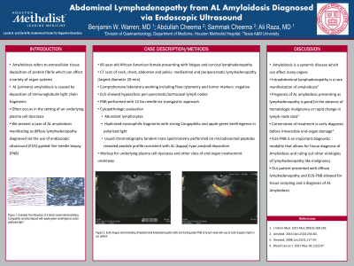
Figure: EUS images demonstrating FNB of an intraabdominal lymph node
Disclosures:
Benjamin Warren indicated no relevant financial relationships.
Abdullah Cheema indicated no relevant financial relationships.
Sammak Cheema indicated no relevant financial relationships.
Ali Raza indicated no relevant financial relationships.
Benjamin Warren, MD1, Abdullah Cheema, 2, Sammak Cheema, 2, Ali Raza, MD1. B0475 - Abdominal Lymphadenopathy from AL Amyloidosis Diagnosed via Endoscopic Ultrasound, ACG 2022 Annual Scientific Meeting Abstracts. Charlotte, NC: American College of Gastroenterology.
