Back


Poster Session B - Monday Morning
Category: Stomach
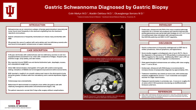
B0720 - Gastric Schwannoma in a Patient With Early Satiety
Monday, October 24, 2022
10:00 AM – 12:00 PM ET
Location: Crown Ballroom

Has Audio

Colin Martyn, MD
Indiana University Internal Medicine Residency - Southwest
Vincennes, IN
Presenting Author(s)
Colin Martyn, MD1, Aladdin Dahbour, MD1, Oluwagbenga Serrano, MD2
1Indiana University Internal Medicine Residency - Southwest, Vincennes, IN; 2Good Samaritan Hospital, Vincennes, IN
Introduction: Schwannomas are an uncommon subtype of benign gastrointestinal mesenchymal tumor found most frequently in the stomach originating from the Auerbach myenteric nerve plexus. Gastric schwannoma is frequently mistreated as it shares many similarities with GIST. We present the case of a patient with early satiety and iron deficiency anemia who was found to have gastric schwannoma on upper endoscopy.
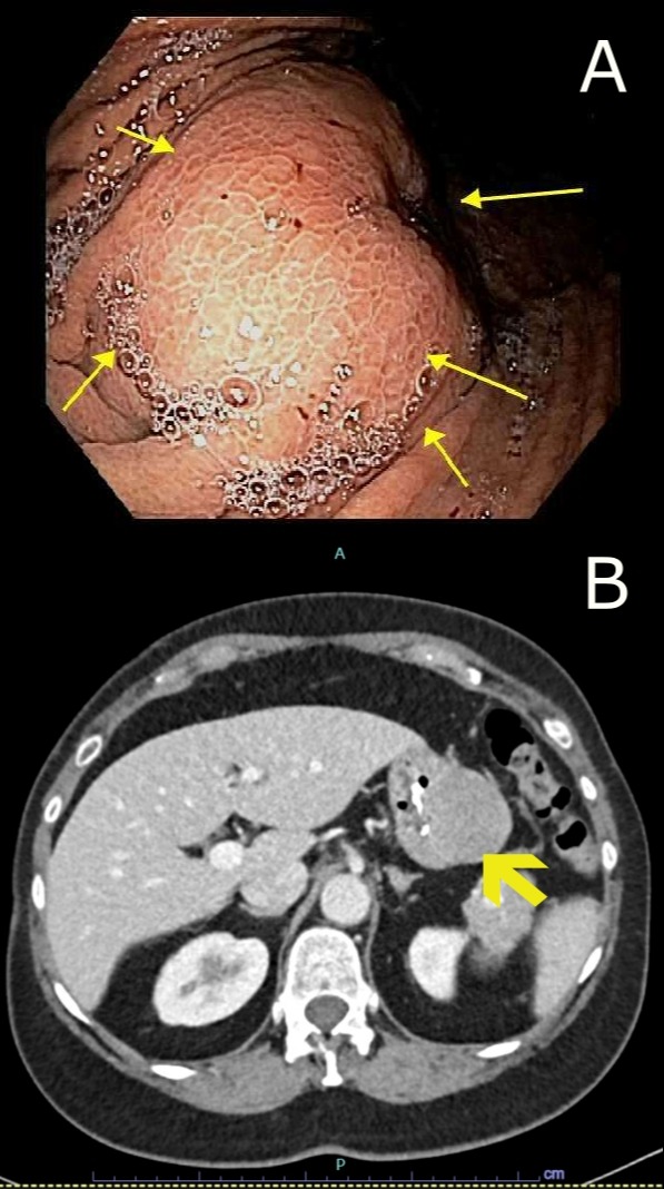
Case Description/Methods: A 59-year-old female with endometriosis and iron deficiency anemia on oral iron supplementation presented to our clinic with complaints of fatigue, frequent post-prandial cough, early satiety, and dark stools. She reported chronic NSAID use and denied abdominal pain, dysphagia, nausea, and unexplained weight loss. Initial CBC demonstrated a hemoglobin of 9.2 g/dL with positive stool guaiac testing. Cessation of NSAIDs led to hemoglobin improvement within normal limits. EGD revealed a roughly 5 cm smooth submucosal mass in the distal gastric body along the greater curvature with firm consistency and a central depression (Figure 1-A). Colonoscopy was unremarkable. Abdominal CT demonstrated an exophytic mass within the gastric wall with relatively homogenous attenuation and enhancement (Figure 1-B). Endoscopic ultrasound with FNA of the mass revealed pathology suspicious for a stromal cell neoplasm and elective laparoscopic partial gastrectomy was performed with findings of a 4.7 x 3.7 x 3.6 cm submucosal mass consistent with schwannoma with Sox10/S100 positivity and DOG-1/c-KIT negativity. The patient resumed a normal diet 2 days after surgery without complications.
Discussion: Gastric schwannoma is frequently misdiagnosed as GIST due to similar prevalence, clinical symptoms, and appearance. Some studies suggest a misdiagnosis rate of up to 96.7%. Key to the diagnosis is the presence of S-100 positivity in schwannoma as GIST is negative for this marker. Additionally, DOG-1 and c-KIT are markers positive in GIST but negative in schwannoma. Most gastrointestinal schwannomas are solitary with sizes ranging from < 1 cm to 28 cm. Endoscopic ultrasound with FNA biopsy is recommended to increase chances of preoperative diagnosis by approximately 10%. Treatment modalities vary based on tumor size, with endoscopic resection suggested for tumors < 3 cm in diameter and surgical excision for larger sized masses. Malignant transformation is extremely rare occurring in roughly 2% of cases with S-100 expression decreasing as dedifferentiated Schwann cells form.

Disclosures:
Colin Martyn, MD1, Aladdin Dahbour, MD1, Oluwagbenga Serrano, MD2. B0720 - Gastric Schwannoma in a Patient With Early Satiety, ACG 2022 Annual Scientific Meeting Abstracts. Charlotte, NC: American College of Gastroenterology.
1Indiana University Internal Medicine Residency - Southwest, Vincennes, IN; 2Good Samaritan Hospital, Vincennes, IN
Introduction: Schwannomas are an uncommon subtype of benign gastrointestinal mesenchymal tumor found most frequently in the stomach originating from the Auerbach myenteric nerve plexus. Gastric schwannoma is frequently mistreated as it shares many similarities with GIST. We present the case of a patient with early satiety and iron deficiency anemia who was found to have gastric schwannoma on upper endoscopy.
Case Description/Methods: A 59-year-old female with endometriosis and iron deficiency anemia on oral iron supplementation presented to our clinic with complaints of fatigue, frequent post-prandial cough, early satiety, and dark stools. She reported chronic NSAID use and denied abdominal pain, dysphagia, nausea, and unexplained weight loss. Initial CBC demonstrated a hemoglobin of 9.2 g/dL with positive stool guaiac testing. Cessation of NSAIDs led to hemoglobin improvement within normal limits. EGD revealed a roughly 5 cm smooth submucosal mass in the distal gastric body along the greater curvature with firm consistency and a central depression (Figure 1-A). Colonoscopy was unremarkable. Abdominal CT demonstrated an exophytic mass within the gastric wall with relatively homogenous attenuation and enhancement (Figure 1-B). Endoscopic ultrasound with FNA of the mass revealed pathology suspicious for a stromal cell neoplasm and elective laparoscopic partial gastrectomy was performed with findings of a 4.7 x 3.7 x 3.6 cm submucosal mass consistent with schwannoma with Sox10/S100 positivity and DOG-1/c-KIT negativity. The patient resumed a normal diet 2 days after surgery without complications.
Discussion: Gastric schwannoma is frequently misdiagnosed as GIST due to similar prevalence, clinical symptoms, and appearance. Some studies suggest a misdiagnosis rate of up to 96.7%. Key to the diagnosis is the presence of S-100 positivity in schwannoma as GIST is negative for this marker. Additionally, DOG-1 and c-KIT are markers positive in GIST but negative in schwannoma. Most gastrointestinal schwannomas are solitary with sizes ranging from < 1 cm to 28 cm. Endoscopic ultrasound with FNA biopsy is recommended to increase chances of preoperative diagnosis by approximately 10%. Treatment modalities vary based on tumor size, with endoscopic resection suggested for tumors < 3 cm in diameter and surgical excision for larger sized masses. Malignant transformation is extremely rare occurring in roughly 2% of cases with S-100 expression decreasing as dedifferentiated Schwann cells form.

Figure: A. Endoscopic appearance of schwannoma as a smooth exophytic submucosal mass with central depression seen along the greater curvature of distal gastric body. B. Abdominal CT demonstrating luminal compression of the gastric body by a schwannoma with relatively homogenous attenuation and enhancement.
Disclosures:
Colin Martyn indicated no relevant financial relationships.
Aladdin Dahbour indicated no relevant financial relationships.
Oluwagbenga Serrano indicated no relevant financial relationships.
Colin Martyn, MD1, Aladdin Dahbour, MD1, Oluwagbenga Serrano, MD2. B0720 - Gastric Schwannoma in a Patient With Early Satiety, ACG 2022 Annual Scientific Meeting Abstracts. Charlotte, NC: American College of Gastroenterology.
